
Evidence date · 2025-05-08 · Research note 2 of 3
1 2 3
See current Track status →
Since the previous research note · Earlier expectation not tested
Track continuity
Since the previous research note: Brain-directed gene delivery protected movement and muscle development in rats with creatine transport deficiency
Previous expectation: Restoring creatine transport along relevant nervous-system pathways might protect movement and muscle development in children if delivery and benefits translate safely.
What this study adds: The May 2025 study examines a different strategy, delivering creatine-synthesis genes to cultured cells with defective transport. It reports increased intracellular creatine in patient-derived fibroblasts, without testing the earlier motor-development expectation.
What changed in this study
For cells to use creatine as an energy buffer, they must either make it or bring it across the cell membrane. In creatine transporter deficiency, the importing route is defective. The disorder is caused by loss-of-function mutations in SLC6A8, the gene for creatine transporter 1, or CT1. It is the most common form of cerebral creatine deficiency syndrome, accounting for about 70% of cases.
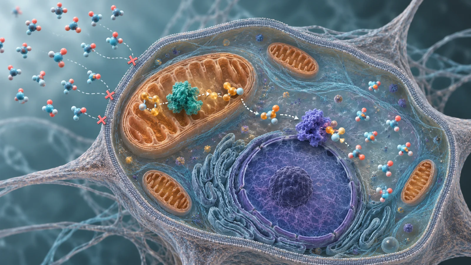
Dietary creatine generally cannot correct the problem when cells lack usable transporter function. A May 2025 laboratory study tested a different workaround: instead of repairing the import route, researchers delivered the genes for two enzymes that make creatine inside the cell. The experiment raised creatine levels in patient-derived skin cells, but its meaning depends on understanding both what those cells demonstrated and what they could not model.
A two-step route around a broken transporter
Creatine helps buffer changing energy demands. Cells can use creatine kinase to move phosphate onto creatine when energy is abundant, creating phosphocreatine, and later transfer that phosphate back to ADP when demand rises. The study asked whether cells unable to import creatine could manufacture enough of it internally to create a usable reserve.
The natural synthesis pathway has two main steps. The GATM gene produces the enzyme AGAT, which combines arginine and glycine to make guanidinoacetate. GAMT then modifies that intermediate, using a methyl donor, to produce creatine. The researchers delivered both GATM and GAMT because increasing only the first step would leave the pathway dependent on whatever GAMT the target cell already possessed.
Their initial experiments supported that reasoning. HEK293T cells had low GAMT expression and no detectable AGAT. Adding AGAT alone increased creatine in one tagged construct, but the increase was much larger when both enzymes were supplied. Co-expression raised intracellular creatine 7.6-fold in the main experiment, compared with empty-vector controls. The result suggested that the complete two-enzyme route could push more material through the pathway than relying on a cell’s limited background machinery.
From a test cell to patient-derived fibroblasts
The researchers next used lentiviral particles to deliver GATM and GAMT into human fibroblasts. Fibroblasts are connective-tissue cells grown from skin samples, so they provide a human patient-derived setting while remaining very different from neurons and other brain cells that would matter for a cerebral disorder.
The comparison included one healthy-control fibroblast line and two CT1-deficient lines carrying different loss-of-function SLC6A8 mutations. The two patient lines came from male donors whose cells had a complete loss-of-function copy of the X-linked transporter gene. This made them useful for testing whether intracellular synthesis could raise creatine without relying on CT1-mediated import.
Before measuring the metabolite, the cells were moved into creatine-free medium supplemented with arginine, glycine and methionine, the precursor materials used by the pathway. The setup reduced the contribution of creatine already present in the medium and supplied the ingredients needed for the introduced enzymes to work. Creatine was measured inside washed cell samples and normalized to total cellular protein, so the reported values describe creatine relative to the amount of cell material analyzed rather than a restoration of tissue-level function.
In healthy-control fibroblasts, co-expression of AGAT and GAMT produced 19.7 nanomoles of creatine per milligram of protein, compared with 1.6 in empty-vector controls. That was a 12.3-fold increase after the cells had grown for about a month without selection. The experiment therefore showed that the introduced pathway could remain active through extended culture and could raise intracellular creatine in a human cell type.
The two patient lines did not respond alike
The patient-derived results were more informative than a single headline number because the two lines behaved differently. In the line carrying the SLC6A8 deletion involving exons 10 and 11, intracellular creatine rose from 1.2 to 4.2 nanomoles per milligram of protein. That is a 3.5-fold increase, but the reported P value was 0.061, just above the study’s stated statistical threshold.
In the second patient line, carrying the SLC6A8 W556X mutation, creatine rose from 0.9 to 10.4 nanomoles per milligram of protein. The resulting 11.6-fold increase met the reported threshold, with P less than 0.05. Each comparison used its own empty-vector control, so the two fold changes should not be read as measurements against one shared baseline.
The important result is therefore not that every patient cell responded by the same amount. The study found that the introduced enzymes could increase intracellular creatine in both disease-derived lines, while the size and statistical strength of the response differed. The experiments do not establish whether the difference arose from the particular SLC6A8 mutations, properties of the cell lines or another feature of the culture system.
The introduced enzymes stayed in place as cells kept growing
A gene-delivery strategy has to do more than produce a biochemical signal briefly. The proteins also need to reach the compartments where the pathway is expected to operate. In the modified fibroblasts, AGAT showed a tubular and punctate pattern consistent with mitochondrial targeting. GAMT was strongly increased throughout the nucleocytoplasmic compartment, matching its expected cellular distribution.
The researchers detected the introduced proteins after several passages without blasticidin selection. In the patient-derived fibroblasts, they also measured cell growth over three days. In standard medium, the modified CT1-deficient lines grew faster than their empty-vector controls, and the same pattern appeared when precursor amino acids were added. The healthy-control fibroblasts showed no meaningful growth difference between the modified and control conditions.
Those observations are useful compatibility checks. They indicate that, in these cultures, the added pathway did not visibly impair cell abundance or short-term growth. They do not provide a quantified long-term expression trajectory, a whole-body safety assessment or evidence that gene delivery would be durable throughout the nervous system. The study also did not compare this synthesis strategy with replacing the defective transporter.
What the creatine measurements leave open
The assay measured total creatine-related material detected by the kit, not the separate proportions of unphosphorylated creatine and phosphocreatine. Because cells contain creatine kinase, some newly synthesized creatine could be converted into phosphocreatine, but the experiments did not determine how much entered either form. A higher total measurement therefore cannot by itself show that the cells gained a larger or more effective energy buffer.
The culture design nevertheless gave the intracellular result a specific meaning. CT1-deficient fibroblasts could not be expected to concentrate creatine through a functioning transporter, and the measurements were taken after exposure to creatine-free medium with precursor amino acids. The researchers also found that the increase in creatine released into the medium was smaller than the intracellular increase. Together, these observations support internal synthesis and retention as the main explanation for the cellular rise under the tested conditions.
A future therapeutic version would face a harder metabolic setting. Cells would need access to enough arginine, glycine, methionine and the methyl donor used by GAMT, while tolerating the demands of running the pathway. The study did not quantify how those resources changed inside the modified cells. It also did not establish whether the same arrangement would be suitable for neurons, which have different energetic demands and cellular environments from fibroblasts.
Why making creatine inside brain cells could matter
The potential value of this approach is conditional: relevant brain cells in people with creatine transporter deficiency might one day be enabled to maintain their own creatine supply even when import through CT1 is faulty. That could provide a way to support the creatine-dependent energy buffering needed for brain function without first restoring transport across the cell membrane.
For that possibility to become a treatment, the delivered genes would have to reach the right cells, remain active at tolerable levels and avoid harmful metabolic consequences. The resulting creatine would also need to participate usefully in cellular energy handling, not merely appear in a total-creatine assay. The May 2025 fibroblast experiments establish a cellular synthesis-bypass branch of the research, not restored brain function, preservation of movement or muscle development, or benefit in treated patients.
Key Sources
- Primary source: Gene delivery of AGAT and GAMT boosts creatine levels in creatine transporter deficiency patient fibroblasts.
DOI or Journal Page · Peer-Reviewed Paper · 2025-05-08