
Evidence date · 2026-01-08 · Research note 3 of 3
1 2 3
See current Track status →
Since the previous research note · Partly supported
Track continuity
Since the previous research note: How a copper-containing scaffold links light-driven chemistry with immune treatment
Previous expectation: A material combining light-triggered tumor injury with immune stimulation might eventually contribute to cancer treatment if its effects proved reproducible and safe.
What this study adds: The January 8, 2026 review describes multiple preclinical frameworks connecting treatment-induced injury with immune agents, including mouse experiments reporting distant tumor suppression. This supports the broader combination strategy while leaving patient benefit and validation of the earlier specific scaffold unresolved.
What changed in this study
Cancer treatment has two related but separate jobs. It can damage tumor cells directly, and it can help the immune system recognize what remains and organize a response. A tumor may release molecular signs of injury without becoming an effective target for immune cells. Conversely, immune stimulation may be too weak if the tumor remains protected by low oxygen, antioxidant defenses, or a physically difficult environment.
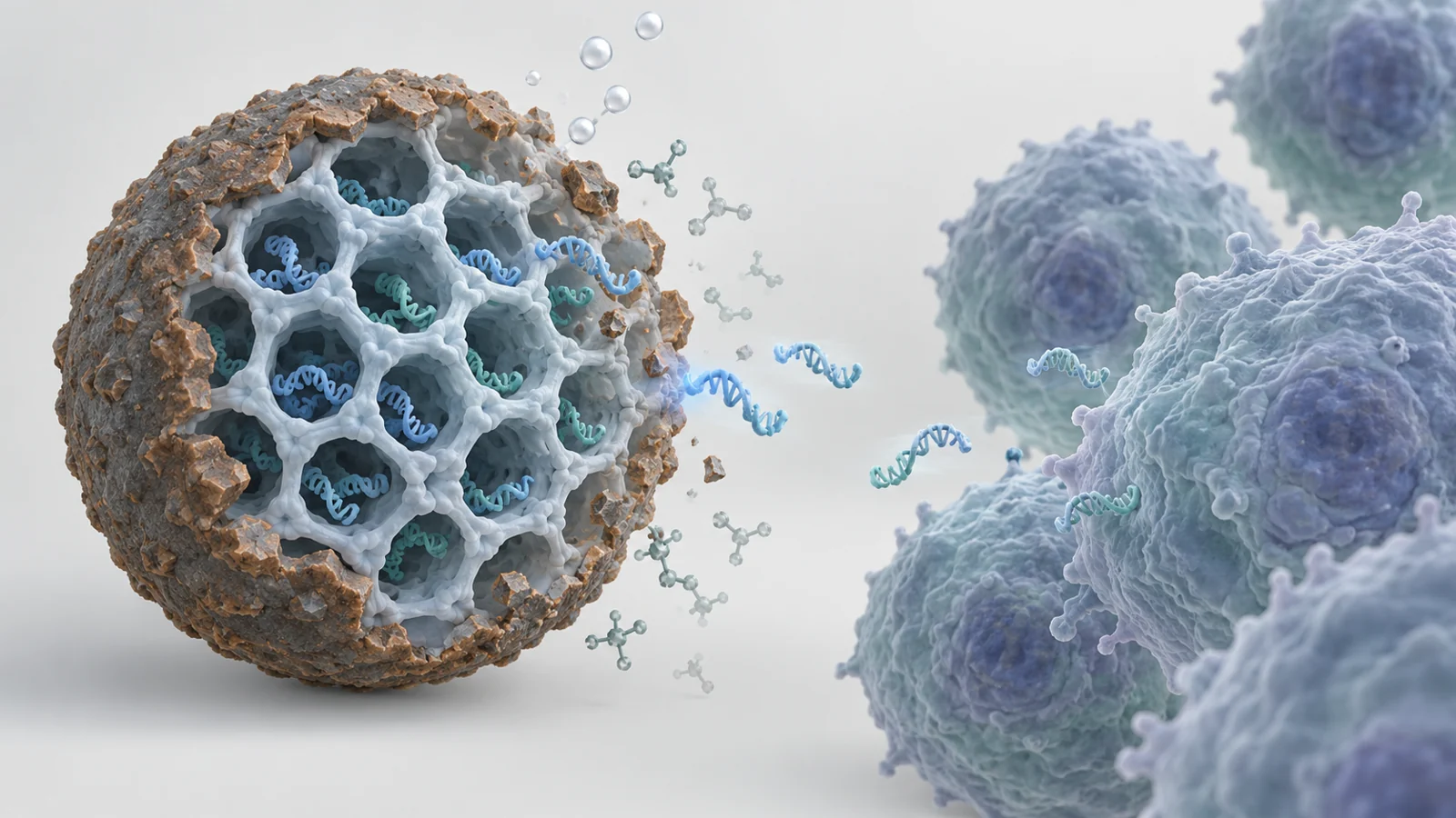
A review published on January 8, 2026, examines how covalent organic frameworks might connect these jobs. COFs are crystalline, porous molecular scaffolds built from lightweight elements joined by strong covalent bonds. Their ordered pores, adjustable surfaces, and modular chemistry allow researchers to use them as treatment sensitizers, chemical modifiers, cargo carriers, or components of imaging-guided systems. The review brings together cell experiments and mouse tumor studies across varied formulations. It does not establish a clinical treatment, a universal response rate, or a ranking of the different designs.
Why tumor destruction and immune response are separate jobs
The central biological idea is immunogenic cell death. This means a form of tumor-cell death that releases antigens and danger signals capable of helping initiate an immune response. Photodynamic therapy, sonodynamic therapy, radiotherapy, and chemodynamic therapy can all be used in COF-based designs to generate damaging reactive molecules or otherwise intensify tumor injury. The resulting signals may give immune cells more information about the tumor, but the signals still need to be amplified and coordinated.
Many tumors are described as immunologically “cold” because they have an immunosuppressive tumor microenvironment and limited T-cell infiltration. Low oxygen can make some treatments less effective. High levels of glutathione can neutralize reactive molecules before they do enough damage. Suppressive immune cells and low baseline tumor immunogenicity add further barriers. These conditions help explain why a framework that merely carries a photosensitizer is not doing the same biological job as a framework that also changes the local chemistry or releases an immune stimulant.
The review therefore treats the material as part of a sequence rather than as a magic particle. A formulation may help create tumor injury, make that injury more visible, deliver a separate immune signal, and provide information about what is happening. Each role has its own measurement and its own failure mode.
A porous scaffold can change tumor chemistry
One example combines several jobs in a single design. Researchers loaded Poly(I:C), an immune-stimulating agent, into a positively charged COF and then grew a manganese-oxide coating over the surface. The resulting composite was described as MnO₂–Poly(I:C)@COF. The porous carrier provides a place for the immune payload, while the manganese oxide changes the chemical conditions around the tumor.
Glutathione is an antioxidant, so high local levels can counter the reactive molecules that photodynamic, sonodynamic, or related treatments are trying to generate. In the reviewed example, the manganese oxide consumed glutathione and helped decompose hydrogen peroxide to produce oxygen. Lowering an antioxidant defense and increasing oxygen availability can create a more favorable setting for reactive-oxygen-based treatment. The composite also released Poly(I:C) and manganese ions to support immune activation, while magnetic resonance imaging was used to monitor the change in the tumor environment.
The point is not that every COF performs these reactions. It is that the scaffold can be engineered so that its carrier function and its chemical function work together. Conventional COFs are often described as metal-free because they are built from light elements, but application-specific designs can incorporate iron, copper, or platinum through suitable binding sites; the manganese-oxide example instead uses a surface coating. The presence of a metal is therefore a design choice in some formulations, not a defining feature of all COFs.
This approach also shows why the immune stimulant cannot be treated as an interchangeable accessory. Oxygen production and glutathione depletion alter the conditions for treatment. Poly(I:C) supplies a separate immune-directed input. The combined design seeks to make tumor injury more likely and to give the immune system additional instructions once injury occurs.
The immune result depends on what is measured
A different example used Fe₃O₄@COF nanoparticles loaded with the CpG immune adjuvant. The iron-oxide core was described as providing chemodynamic activity and magnetic properties, while the COF component supported sonodynamic treatment. In mice, the formulation suppressed tumor growth under ultrasound-associated treatment. When CpG was included, the treatment produced stronger tumor inhibition than the sonodynamic treatment alone in the reported comparison.
The same study also measured dendritic-cell maturation in tumors. That result matters for a different reason. Tumor growth is an outcome at the level of the lesion. Dendritic-cell maturation is a cellular indication that the immune environment has changed in a direction relevant to immune coordination. The review reports that both CpG and sonodynamic treatment increased the proportion of mature dendritic cells, providing an immune-related measurement alongside tumor growth rather than treating tumor shrinkage as proof of immune activation.
This distinction keeps the evidence in proportion. A mouse tumor measurement can show that a formulation affected tumor progression under the tested conditions. A dendritic-cell measurement can show that immune-associated biology changed. Neither measurement by itself establishes that people would develop durable protection, that treatment-resistant tumors would respond, or that the same formulation would be safe outside the experimental setting.
Activation has to reach the material
A carefully designed photosensitizer is useful only where the activating light can reach it. The review describes COF-606, which absorbed two-photon excitation at 808 nanometers. Under the reported imaging conditions, one-photon excitation at 560 nanometers reached an imaging depth of 130 micrometers, whereas two-photon excitation reached 233 micrometers. Those numbers make the physical problem concrete: the comparison concerns imaging depth in micrometers, not a demonstrated treatment depth in a human tumor.
The review also reports tumor suppression and increased levels of ATP, calreticulin, and HMGB1 after treatment with COF-606 and 808-nanometer irradiation. These observations connect the material’s activation to markers associated with immunogenic cell death, but the imaging comparison should not be converted into a claim that two-photon treatment can reach any clinically relevant tumor depth.
Ultrasound offers a different route for activating a material in deeper lesions. The reviewed Pg@Fe-COF design used iron coordination to support glutathione depletion and chemodynamic activity while enabling sonodynamic treatment. The review notes that combining this approach with an anti-PD-L1 antibody produced stronger therapeutic outcomes in the reported experiments. Ultrasound is not universally better than light; it changes the physical access problem and avoids the cutaneous photosensitivity concern associated with some light-activated treatments. The appropriate activation route depends on the formulation, the tumor location, the energy source, and the control available over exposure.
Local immune architecture is not the same as distant control
Some of the most interesting findings concern what happens beyond the initially treated cells. The review describes COF-based phototherapy systems that promoted tertiary lymphoid structures, or TLS, within tumors. These are lymph-node-like immune hubs formed locally in the tumor environment. In the reported work, inflammatory signaling was associated with the maturation and recruitment of T cells and B cells, creating a more organized immune setting inside the lesion.
Separate reviewed experiments reported suppression of a distant tumor. One multifunctional COF design combined photodynamic, photothermal, and chemodynamic effects and was reported to inhibit both primary and distant tumors in mice. Other platforms used different structural or biological modifications. These findings address a different question from TLS formation: they ask whether treatment of one site is associated with a measurable effect at another site.
Both observations are important, but they should not be merged into a single endpoint. TLS formation describes local immune organization. Distant tumor suppression describes an effect measured beyond the treated lesion. Neither observation alone demonstrates durable immune memory, prevention of recurrence, cure, or a reliable abscopal response in patients. The value of the review is to show how these possibilities arise from different experimental designs, not to declare that all COFs produce them.
Translation turns durability into a design problem
The material must remain functional long enough to reach the tumor, respond to its activating conditions, and release any therapeutic cargo at the intended time. It may also need to break down or clear appropriately afterward. Those requirements can pull in opposite directions. Some imine- or boronate-ester-linked COFs may be vulnerable to acidic lysosomal conditions or oxidative stress. That degradation could compromise structural performance, but controlled disassembly might also help release a loaded agent. The useful question is therefore not simply whether a framework is stable, but whether its persistence and breakdown occur at the right place and time.
Control is also needed at the level of activation. A photosensitizer can cause nonspecific phototoxicity when light dosing is poorly controlled or healthy tissue is unintentionally exposed to irradiation. Porphyrin-based components may undergo photobleaching, reducing phototherapeutic performance. Even a framework with favorable laboratory behavior would need its biodistribution, long-term metabolic fate, immune compatibility, and clearance to be understood before its risks could be judged in people.
Manufacturing creates a separate bottleneck. A formulation that works in a carefully controlled mouse experiment may still be difficult to produce with consistent particle properties, loading, coating, purity, and stability at larger scale. Reproducible production is not a cosmetic improvement; it is part of making the biological result testable from one batch, study, or clinical setting to the next. The review points toward biodegradable building blocks, stimulus-responsive linkages, greener synthesis, continuous-flow or microwave-assisted production, and biomimetic coatings as possible directions, while emphasizing the need for coordinated work among materials scientists, pharmacologists, immunologists, and clinicians.
Why this could matter for tumors that resist immunotherapy.
A locally activated COF treatment could become useful against difficult tumors if it can both intensify injury at the treated site and recruit immune activity capable of acting beyond that site. For a patient whose tumor responds poorly to checkpoint blockade, the potential benefit would be more than a carrier delivering one drug: it would be a controllable way to connect local treatment with a broader immune response.
That possibility remains conditional from the January 8, 2026 perspective. The mouse findings would need to translate into reliable systemic responses in people, supported by safe delivery, controllable activation, appropriate persistence and breakdown, and reproducible formulations. The review shows why porous molecular scaffolds are being explored for this purpose; it does not show that a COF-based cancer treatment has crossed that clinical boundary.
Key Sources
- Primary source: Covalent organic frameworks for cancer immunotherapy: mechanisms, applications, and prospects.
DOI or Journal Page · Review or Meta-Analysis · 2026-01-08