
Evidence date · 2023-04-06 · Research note 1 of 3
1 2 3
See current Track status →
On April 6, 2023, researchers reported a way to combine two functions inside the same cancer-fighting cell. Their engineered cells were designed to recognize HER2, a surface protein found at high levels on some breast-cancer cells, while also releasing soluble programmed cell death protein 1, or soluble PD-1. The study tested the cells in short laboratory killing assays and in mice carrying breast tumors, including tumors made from a trastuzumab-resistant human cancer cell line.
The central question was practical. If an immune cell is already being directed toward a tumor, could it also release a protein that helps counter local immune inhibition? The results suggested an advantage for that combined design over HER2-directed cells alone, and over HER2-directed cells accompanied by separately injected soluble PD-1. They remained preclinical findings from engineered cell and mouse models, rather than evidence of benefit for patients.
Two jobs built into one immune cell
The researchers used NK-92, a laboratory natural killer cell line, as the starting material. Natural killer cells can damage abnormal cells, but the engineered version added a chimeric antigen receptor, or CAR. In this study, the receptor used a HER2-specific binding component to help the cells recognize target cells carrying HER2, then included intracellular signaling elements intended to activate the NK cell after recognition.
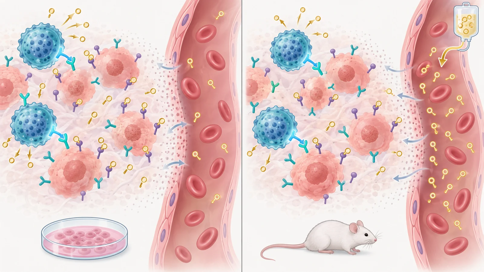
The experimental cells added another component to that receptor design. They were made to release the extracellular portion of PD-1 as soluble PD-1, rather than keeping the protein attached to the cell surface. Soluble PD-1 is a released protein, not an anti-PD-1 antibody. Its intended role here was to interfere with the interaction between PD-1-related inhibitory signaling and PD-L1, a ligand found on many cancer cells and in the tumor environment. The proposed arrangement therefore gave one cell two distinct tasks: find HER2-bearing targets and supply a local blocker of an inhibitory interaction.
The construct produced soluble PD-1 in culture. The researchers detected a large amount in the culture fluid from the engineered cells, while the protein was not detected in the cell lysate in the same experiment. That result supported the intended release behavior before the team compared cancer-cell killing.
The clearest added activity was against JIMT-1 cells
The laboratory experiments used three breast-cancer cell lines to separate recognition from inhibitory signaling. MDA-MB-231 cells had very low HER2 and high PD-L1. MCF-7 cells had high HER2 and low PD-L1. JIMT-1 cells had high levels of both HER2 and PD-L1 and came from a patient-derived trastuzumab-resistant HER2-positive breast cancer line.
HER2-CAR-NK cells were more active against the HER2-rich MCF-7 and JIMT-1 cells than unmodified NK-92 cells. The cells that also released soluble PD-1 showed greater apparent killing than HER2-CAR-NK cells against all three tested lines, with the clearest added activity against JIMT-1. At an effector-to-target ratio of 10 to 1, the reported killing of JIMT-1 cells by the secreting cells exceeded 80 percent.
Those results fit the study’s two-part design without proving that either marker alone determined the response. HER2 gave the engineered cells a recognition target, while PD-L1 provided a setting in which blocking an inhibitory interaction might matter. The assay measured cancer-cell damage after six hours under controlled culture conditions. It did not reproduce the full structure, circulation, or immune environment of a human tumor.
Mouse tests separated the model questions
The first animal setting used EMT6 mammary tumor cells modified to express human HER2 in immunocompetent BALB/c mice. This model allowed the researchers to ask whether the engineered cells could slow the growth of a HER2-bearing tumor in a mouse with an intact mouse immune system. Once tumors had formed, mice received parental NK-92 cells, HER2-CAR-NK cells, or the cells that also secreted soluble PD-1. The engineered groups were treated through the tail vein on a recurring schedule, with interleukin-2 used to help maintain immune-cell activity.
HER2-CAR-NK cells slowed tumor growth more than parental NK-92 cells, while the soluble-PD-1-secreting cells produced a greater delay than HER2-CAR-NK cells in the reported comparison. Tumor weight showed a similar pattern. One described BALB/c experiment used three mice per group, a small number that makes the result useful as an experimental signal but unsuitable for estimating how consistently the effect would appear in a broader population.
The second setting addressed a different question. The researchers implanted human JIMT-1 tumors, the trastuzumab-resistant line used in the culture experiments, into NOG mice that had been given human immune cells. This extended the test from a mouse tumor engineered to display human HER2 to a human resistant breast-cancer cell line in a mouse host containing human immune components. In that model, the soluble-PD-1-secreting cells again showed the strongest reported inhibition of tumor growth among the compared NK-cell treatments, and soluble PD-1 was detected in both the blood and tumor tissue after treatment.
Secreting cells outperformed the separate-protein combination in mice
The most informative test compared two ways of providing the same general combination. One group received cells engineered to recognize HER2 and release soluble PD-1. Another received HER2-CAR-NK cells plus soluble PD-1 protein injected separately. For the separate-treatment comparison, the authors used soluble PD-1 at 2.4 ng/mL, a concentration they related to protein measured in culture supernatant from 6 million secreting cells.
The engineered secreting cells controlled the tumors more strongly than the combination of HER2-CAR-NK cells and separately administered soluble PD-1. The protein measurements offered a possible explanation for why delivery route might matter. The separate-protein combination produced higher soluble-PD-1 levels in serum but lower levels in tumor tissue, whereas the secreting-cell treatment showed more soluble PD-1 in the tumor tissue. In the humanized JIMT-1 setting, soluble PD-1 was described as heavily distributed in tumors treated with the secreting cells.
That pattern is consistent with the idea that a HER2-directed cell can carry its added protein-producing function toward a tumor, rather than relying on a protein injected throughout the body to reach the same location. It remains an interpretation of the observed distribution and tumor-control results. The treatment schedules and protein measurements do not establish equivalent systemic exposure, and the measurements alone do not prove that tumor accumulation was the sole reason for the efficacy difference.
What the animal readouts can and cannot show
The study found no significant changes in body weight or temperature among the groups receiving the engineered NK cells, and it found no apparent injury in the examined heart, liver, spleen, lung, or kidney tissues during the experiments. These observations provide bounded tolerability information for the tested mouse settings. They do not establish freedom from serious toxicity, human safety, or a safety advantage over CAR-T cells or approved checkpoint treatments.
The models also impose important limits on interpretation. NK-92 is a cell line rather than a finished human therapeutic product. The tumors were implanted or engineered experimental systems, and the humanized mouse model cannot reproduce every feature of a patient’s tumor. The treatment also relied on interleukin-2 to support immune-cell activity. The supplied findings do not establish a clinical HER2 or PD-L1 threshold for choosing patients, overcome every mechanism of trastuzumab resistance, or show durable remission, survival improvement, manufacturing readiness, or treatment availability.
What this could mean for resistant breast cancer
The study pointed toward a possible cell therapy for patients whose HER2-positive tumors no longer respond adequately to trastuzumab. In that future use, one engineered immune cell could combine direct recognition of HER2-bearing cancer cells with local release of a protein intended to ease nearby immune inhibition. The tumor-versus-serum comparison in the EMT6-hHER2 model supports interest in the delivery concept, while the separate JIMT-1 experiment extends the tumor-control finding to a trastuzumab-resistant cell line.
Whether the idea becomes clinically useful would depend on the same advantages translating into durable tumor control with acceptable safety in people. The mouse results support further development of the combined design, but they do not promise that it will replace existing treatment or benefit patients until its behavior, risks, and effectiveness are established in human studies.
Key Sources
- Primary source: Engineering a HER2-CAR-NK Cell Secreting Soluble Programmed Cell Death Protein with Superior Antitumor Efficacy.
DOI or Journal Page · Peer-Reviewed Paper · 2023-04-06