
Evidence date · 2026-02-01 · Research note 3 of 3
1 2 3
See current Track status →
Since the previous research note · Earlier expectation not tested
Track continuity
Since the previous research note: How an overactive arousal circuit shaped sleep in amyloid-model mice
Previous expectation: Safely reducing excessive arousal could help people with early Alzheimer's-related sleep disruption sustain sleep if the mouse pathway also operates in humans and benefits endure.
What this study adds: This study adds a human cross-sectional association between an MRI proxy of locus coeruleus integrity and slow sleep rhythms, with exploratory sex and regional vascular findings. It does not test excessive firing, the proposed feedback mechanism, or a treatment that reduces arousal.
What changed in this study
Sleep is often summarized by asking how long someone spends in a deep stage. But the brain’s electrical activity during that stage can vary too. Are stronger slow sleep waves related to the health of a particular brain region, even when the percentage of time assigned to the deepest stage does not show the same pattern?
A study published on February 1, 2026, examined that question in 58 adults aged 50 to 80. The group included 11 cognitively healthy controls, 30 people with mild cognitive impairment due to Alzheimer’s disease, and 17 people with Alzheimer’s dementia. The researchers combined overnight polysomnography, an MRI measure of the locus coeruleus, ratings of small spaces around blood vessels, and cerebrospinal-fluid measurements of noradrenaline.
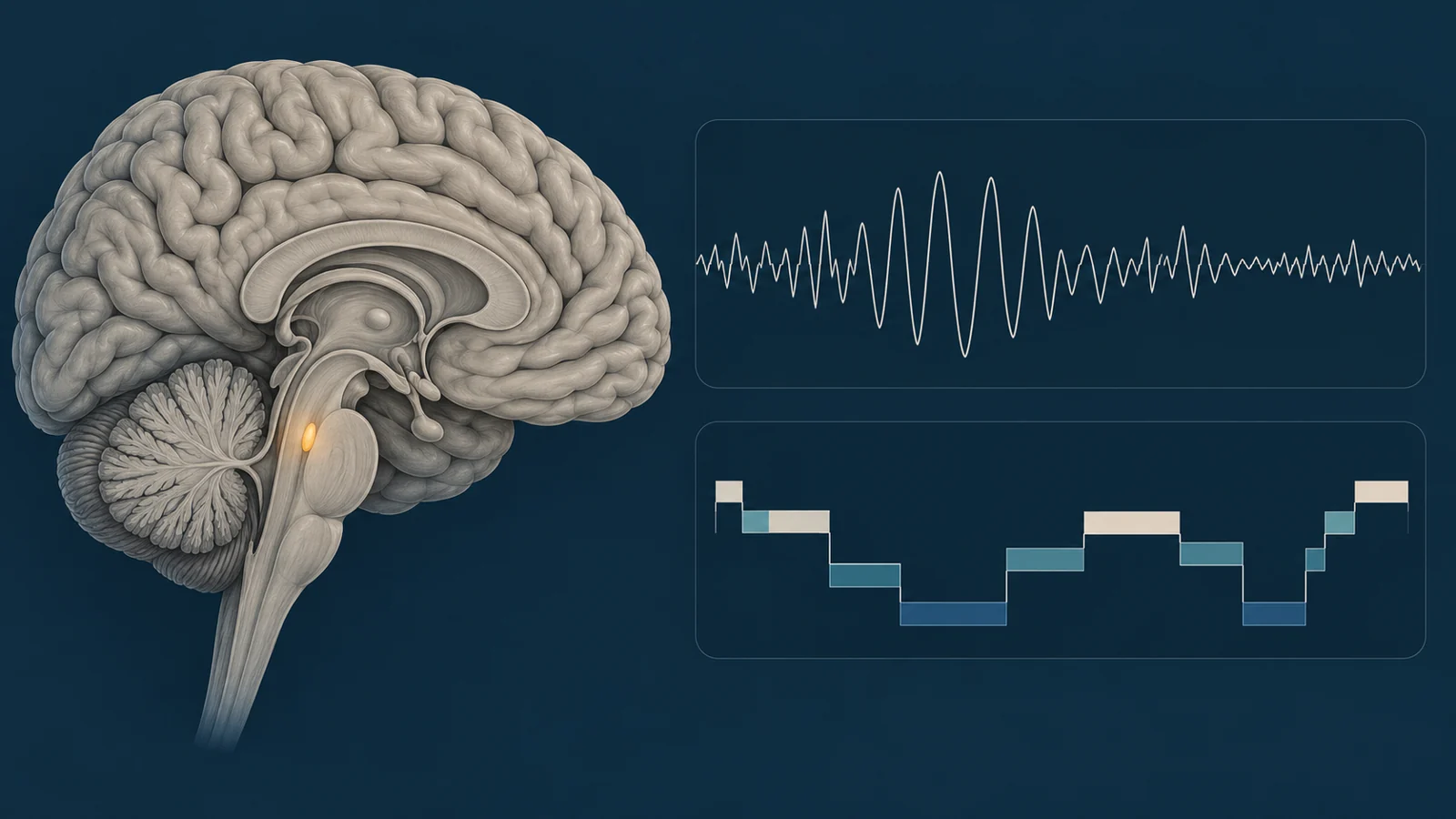
The locus coeruleus is a small brainstem region involved in arousal, attention, and sleep–wake regulation. It is also an early site of Alzheimer’s-related change, according to the paper’s background. In this study, the question was not whether the locus coeruleus explained every sleep difference between diagnostic groups. It was whether individual variation in an MRI-derived measure of its integrity tracked variation in slow sleep rhythms across the group.
Deep sleep has more than one measurable feature
The researchers looked at electrical activity during N2 and N3 sleep using frontal electroencephalography recordings. They calculated power in several frequency ranges. Slow oscillations covered 0.5 to 1 hertz, while the broader slow-wave activity range covered 0.5 to 4 hertz. Delta activity occupied the 1 to 4 hertz range.
These measurements describe the strength of recurring electrical rhythms. They are not the same as the percentage of the night assigned to a sleep stage. A sleep-stage score answers how much time was classified as N3, while spectral power estimates how prominent particular rhythms were during the analyzed periods of N2 and N3. Two people could therefore receive similar stage-based descriptions while showing different slow-wave power.
The MRI measure also required interpretation. It did not directly count locus coeruleus neurons or record their moment-to-moment activity. Instead, the researchers estimated locus coeruleus integrity from MRI signal in the region compared with a reference area. It was an indirect structural measure of the brainstem region, paired with a physiological measure of sleep rhythms.
The adjusted analysis connected brainstem integrity with wave strength
After accounting for demographics, disease stage, and prescribed medication use, higher locus coeruleus integrity was associated with greater slow-wave activity and greater slow-oscillation power. The association was positive for both measures, suggesting that participants with a stronger MRI-derived integrity measure tended, on average, to show more power in the slow rhythms recorded during sleep.
The pattern was not identical across every sleep measure. The association with delta power was weaker and did not reach statistical significance in the interaction model. Locus coeruleus integrity was also not significantly associated with the percentage of sleep spent in N3. The result therefore concerns the electrical character of slow sleep activity more clearly than the amount of time assigned to the deepest stage.
The distinction matters because the study does not show that locus coeruleus integrity accounts for the full difference in sleep between healthy controls, people with mild cognitive impairment, and people with Alzheimer’s dementia. The researchers did not detect significant group differences in locus coeruleus integrity in this relatively small sample. Their interpretation is more specific: across cognitively healthy adults and people along the Alzheimer’s continuum, individual differences in the brainstem measure were related to individual differences in slow-wave physiology.
Sex and vascular findings remain exploratory
The locus-coeruleus relationship was stronger in women than in men for slow-oscillation and broad slow-wave power. The sex interaction was statistically significant for those measures, but the authors describe analyses involving sex as exploratory because the sample was modest. The finding does not establish a hormonal explanation, nor does it show that women and men should receive different treatments.
The study also examined perivascular spaces, small spaces visible around blood vessels on MRI. These were rated separately in the basal ganglia and the centrum semiovale using a visual scale. The ratings describe anatomy in those regions; they do not directly measure the movement of brain fluid or the removal of amyloid or tau.
In reduced regression models, greater basal-ganglia perivascular-space burden was associated with lower slow-wave and slow-oscillation power. The corresponding associations for perivascular spaces in the centrum semiovale were not statistically significant. Exploratory tests of whether vascular burden changed the locus-coeruleus association produced trends, but the proposed interactions did not reach statistical significance.
The exploratory analyses identified a stronger locus-coeruleus association in women and an association between basal-ganglia perivascular-space burden and lower slow-wave power. They did not establish that perivascular-space burden modified the locus-coeruleus association. How these findings fit together biologically remains unresolved.
Why the MRI measure and noradrenaline result differed
Cerebrospinal-fluid noradrenaline levels were not associated with the sleep parameters measured in the study. At first glance, that may seem difficult to reconcile with the relationship between locus coeruleus integrity and slow-wave power, because the locus coeruleus is a noradrenergic brainstem nucleus.
The authors propose one possible explanation. A cerebrospinal-fluid noradrenaline measurement is a relatively static snapshot. It may not capture the changing noradrenergic activity that occurs across sleep and wake states. An MRI-derived integrity measure may instead reflect the longer-term condition or capacity of the locus coeruleus, which could relate more closely to the organization of sleep rhythms. This is a proposed interpretation, not a demonstration that one measure is generally superior to the other.
The same caution applies to the perivascular-space findings. The spaces are an anatomical feature seen on MRI, not a direct test of glymphatic function or brain-fluid clearance. The researchers did not directly measure glymphatic activity, so the study cannot turn the vascular association into a claim about how efficiently the brain removes particular proteins during sleep.
What the study can and cannot establish
This was a single-center, cross-sectional observational study. The measurements were taken as a snapshot rather than during a long-term follow-up. As a result, the direction of the relationship remains open. Locus coeruleus changes could contribute to altered sleep rhythms, sleep disruption could contribute to brain changes, or other factors could influence both.
The sample size also limits what can be learned from subgroup comparisons and interaction tests. The sex-related and vascular findings are useful for generating hypotheses, but they need confirmation in larger and more diverse groups. The MRI measure is an indirect proxy and can be affected by scanner and sequence characteristics, movement, registration, and segmentation. Visual ratings of perivascular spaces also introduce measurement variability. Adjusting for prescribed medications reduces one source of confounding but cannot remove every influence of psychotropic medication or other unmeasured factors.
The MRI association should not be read as evidence of excessive locus coeruleus firing, and it does not confirm a specific adrenergic feedback mechanism or potassium-current pathway. Nor does it provide an individual diagnostic or prognostic threshold. A nonsignificant result, such as the absence of a detected association with N3 percentage or cerebrospinal-fluid noradrenaline, does not prove that no relationship exists; it means that this study did not establish one under its measurements and statistical power.
Most importantly, the researchers did not test a treatment. The findings do not show that changing locus coeruleus signaling improves sleep, memory, daily functioning, or the course of Alzheimer’s disease. They identify a relationship worth testing more directly, not a clinical rule or an intervention that is ready to use.
What this could change
Older adults and people living with Alzheimer’s could eventually benefit from treatments designed to preserve the brain’s slow sleep rhythms, if future longitudinal and mechanistic studies show that the locus coeruleus contributes causally to their decline and that the pathway can be modified safely. Such treatments might aim to support the relevant brainstem signaling system or protect the conditions that allow restorative sleep activity to persist.
That possibility depends on more than reproducing an MRI association. Researchers would need to show that an intervention changes the targeted biology, produces a sustained improvement in slow-wave physiology, and delivers meaningful benefits for people rather than only changing a laboratory measure. Any prospect of preserving cognition or slowing Alzheimer’s progression would be further downstream and remains unproven by this study.
Key Sources
- Primary source: Relationship between locus coeruleus and slow-wave sleep in aging and Alzheimer's disease.
DOI or Journal Page · Peer-Reviewed Paper · 2026-02-01