
Evidence date · 2022-01-21 · Research note 1 of 3
1 2 3
See current Track status →
A brain system that helps keep us awake might seem like an unlikely place to look for clues about restorative sleep. Yet the locus coeruleus, a small paired center in the brainstem, is active at carefully timed points across the sleep-wake cycle. Its main chemical messenger, norepinephrine, reaches much of the brain, including areas involved in memory, emotion and sensory processing.
A review published on January 21, 2022, examines how this system participates in wakefulness, sleep stages and brief events within sleep. It also considers why the locus coeruleus has become relevant to Alzheimer’s disease research. The interest is not based on one decisive human finding. It comes from several kinds of evidence that answer different questions: experiments that manipulate the system in animals, observations of human brain tissue after death, and newer human imaging studies that associate locus coeruleus measures with sleep or cognitive outcomes.
An alerting center that works within a larger circuit
The locus coeruleus is located in the dorsal part of the rostral pons, beside the fourth ventricle. Despite its small size and relatively small number of neurons, it is the central nervous system’s primary source of norepinephrine. Its neurons have widely branching axons, allowing norepinephrine to influence distant regions such as the hippocampus, amygdala, thalamus and neocortex.
That reach helps explain both its importance and its limits. The locus coeruleus is part of an ascending arousal system that also includes brainstem nuclei releasing acetylcholine and serotonin, the ventral tegmental area releasing dopamine, the basal forebrain, and hypothalamic systems involving histamine and orexin. Sleep-promoting circuits in the preoptic area and other regions counterbalance these wake-promoting influences.
The result is a network, not an isolated on-off switch. Early lesion studies produced inconsistent effects on the amount of time animals spent awake or asleep. Some of that variation may reflect how completely the locus coeruleus was damaged and how surviving neurons compensated. Even extensive, more selective damage in later experiments did not necessarily eliminate total daily wakefulness, but it weakened the ability of a novel environment to sustain wakefulness. The system can therefore be crucial for particular demands or transitions without determining every hour of the day by itself.
Awakenings reveal the system’s leverage
More precise methods made it possible to test what happens when locus coeruleus neurons are activated or inhibited at specific moments. In animal experiments reviewed in the paper, light-based inhibition during the active period reduced wakefulness and increased transitions from waking into sleep. Activating the neurons during the inactive period produced immediate transitions from sleep to wakefulness.
The same system also influenced awakenings caused by the outside world. After activation of locus coeruleus neurons, rats were more likely to wake in response to an auditory stimulus; inhibition reduced that probability. The findings point to a role in both internally generated transitions and responses to sensory disturbance.
The chemical messenger mattered as well as the neurons themselves. In a mouse experiment, selectively disrupting an enzyme needed to make norepinephrine reduced the duration of wakefulness and abolished the immediate sleep-to-wake response produced by stimulating locus coeruleus neurons. Some locus coeruleus neurons can produce or release other compounds, so this result helped separate the contribution of norepinephrine from the broader activity of the cells.
These experiments show strong leverage over waking transitions, but the lesion findings add an important qualification. A system can help open the gate to wakefulness while operating alongside other arousal circuits that can compensate when part of the network is damaged. The biology is better described as coordinated control of transitions than as a single switch that dictates the entire sleep budget.
Sleep is a sequence, not just a total
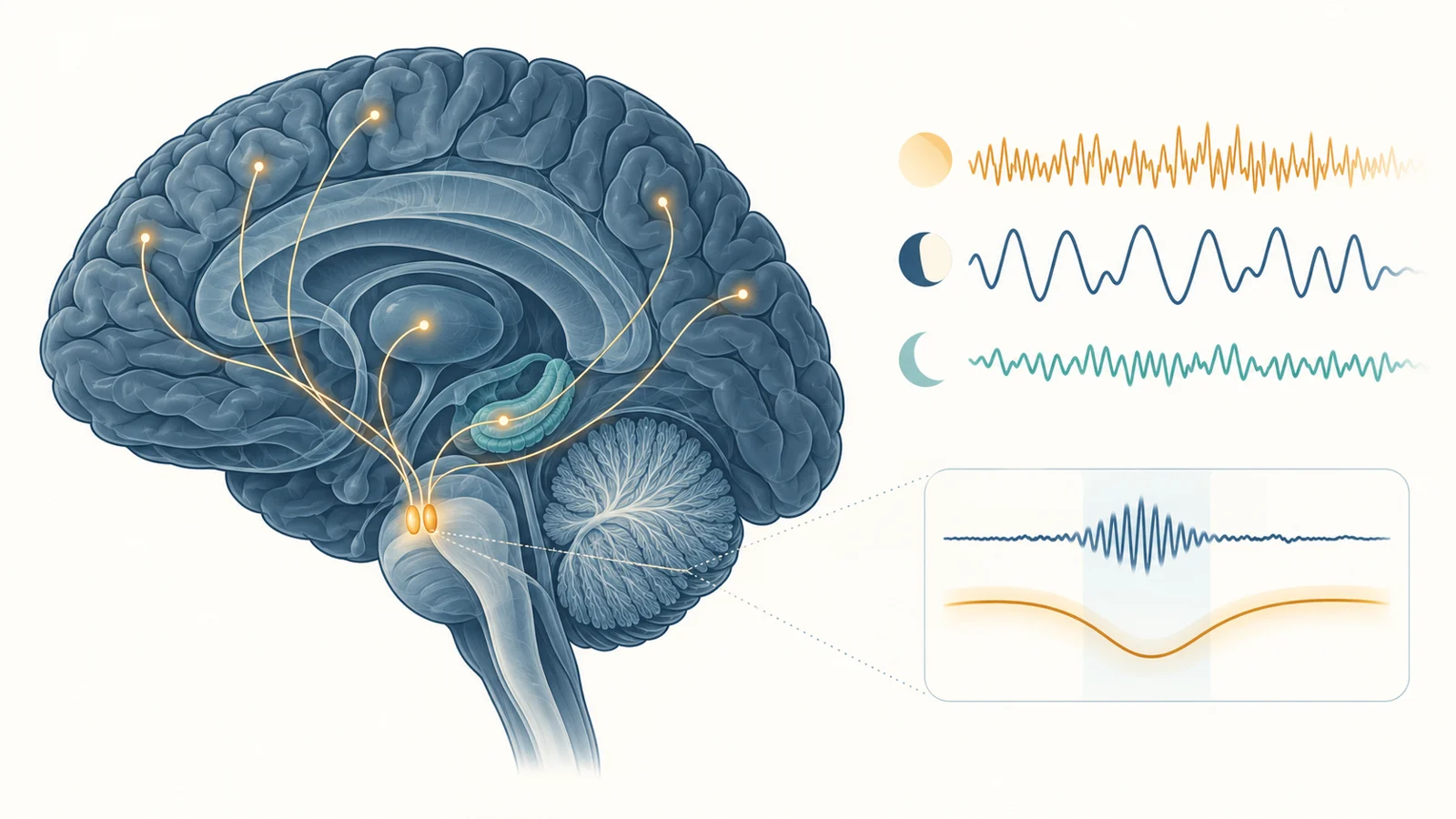
The timing of locus coeruleus activity becomes clearer when sleep is considered as a sequence of states and events rather than a single number of hours. During wakefulness, its neurons are generally active. Their activity falls during non-REM sleep and reaches its lowest level during REM sleep, when locus coeruleus neurons are described as nearly silent. Earlier models proposed a reciprocal relationship between REM-off norepinephrine neurons and REM-on cholinergic neurons in the brainstem, although the wider circuit controlling REM sleep remains an active area of investigation.
Non-REM sleep contains its own fine structure. Sleep spindles are brief bursts of rhythmic brain activity, commonly described as trains of 12–16 Hz waves. In the experiments discussed by the review, locus coeruleus activity dropped in the seconds before a spindle, rose during the spindle and fell again afterward. More recent rodent work linked troughs in extracellular norepinephrine to spindle activity.
Manipulating the system changed this pattern. Stimulating locus coeruleus neurons reduced spindle occurrence and interfered with sleep-dependent memory consolidation. Inhibiting them increased spindle density but also disturbed the timing of spindles. Other recordings found that locus coeruleus activity during non-REM sleep could change with learning and align with the rising phase of slow cortical oscillations.
This makes the question more precise than whether a person is simply awake or asleep. Norepinephrine may help organize when brief sleep events occur and how they are coordinated with other brain rhythms. These findings raise a question for intervention research: how would increasing or suppressing this system affect its different functions across sleep stages and brief sleep events?
Why Alzheimer’s enters the picture
The Alzheimer’s connection begins with two observations that should be kept distinct. Human post-mortem studies have found that the locus coeruleus is among the earliest sites where abnormal tau pathology appears across the lifespan. The review cites post-mortem evidence of abnormally phosphorylated tau in the locus coeruleus in most individuals by age 40. Substantial loss of locus coeruleus neurons, however, has been described as a feature associated with Alzheimer’s disease rather than a general consequence shared equally by other tau-related disorders.
A second line of evidence comes from animal models. Sleep and wakefulness regulate the release and clearance dynamics of amyloid-beta and tau, while Alzheimer’s-related pathology can itself disrupt sleep-wake patterns. In mice, repeated sleep disruption was associated with marked changes in locus coeruleus neurons and their axonal projections. In a mouse model of tauopathy, repeated shortened sleep accelerated tau accumulation within locus coeruleus neurons, its spread to connected regions, and the appearance of neurobehavioral problems. Some structural and cognitive effects remained long after the sleep-disruption protocol ended.
These observations motivate a reinforcing model: sleep-wake disruption could stress the locus coeruleus, while pathology in the system could further disturb sleep organization. But the mouse exposures do not provide a human threshold for harmful sleep loss, and the animal sequence cannot be treated as an established timeline for people. The human post-mortem observation also shows where pathology can appear; by itself, it does not determine whether sleep changes, tau accumulation or neuronal loss comes first in an individual.
What human imaging can connect
Studying the locus coeruleus in living people was difficult because the structure is small and deep in the brainstem. Advances in MRI now make it possible to estimate aspects of its structural and functional integrity, particularly with high-field methods. The resulting MRI contrast is a proxy thought to reflect neuronal and fiber projection density; it is not a direct count of living locus coeruleus neurons.
The review describes a gradual decline in MRI-assessed locus coeruleus contrast beginning around the fifth decade in cognitively unimpaired older adults. Contrast was also lower in people with Alzheimer’s disease and associated with amyloid-beta pathology, while another study connected lower contrast with tau pathology in older adults without cognitive symptoms. These findings make the system measurable across aging, but they do not turn one scan into a diagnosis.
Two human examples illustrate the kind of evidence now available. In 481 older men from the Vietnam Era Twin Study of Aging, lower locus coeruleus contrast was associated with worse self-reported daytime functioning. Lower structural integrity and greater daytime dysfunction each predicted higher odds of later conversion to mild cognitive impairment. The outcome involved daytime function and cognitive status in a relatively large aging cohort.
A separate study of 72 cognitively unimpaired older adults examined nighttime awakenings. Lower integrity in the middle-to-caudal locus coeruleus was associated with more frequent self-reported nocturnal awakenings, particularly among people with higher plasma total tau. This was a smaller group, and the measured outcome was a reported sleep feature rather than conversion to mild cognitive impairment.
Both studies are associations. They connect indirect brain measures with reported functioning, sleep or later cognitive outcomes, but they do not establish the temporal order linking sleep disruption, locus coeruleus alteration and cognitive decline. Fragmented sleep alone is not evidence that a person has Alzheimer’s disease or damage in this brainstem system.
The causal order remains open
The next challenge is to follow the same people repeatedly. Combining locus coeruleus MRI, objective sleep measures such as EEG or actigraphy, sleep reports and cognitive assessments could show whether changes in the system precede sleep disruption, follow it, or develop alongside another process. Such studies would also need to account for factors such as depression and anxiety, which can relate to both locus coeruleus function and sleep quality.
The locus coeruleus should be tested against other arousal systems affected in Alzheimer’s disease, including orexin-producing cells in the lateral hypothalamus and histaminergic neurons in the tuberomammillary nucleus. Because orexin neurons send strong inputs to the locus coeruleus, changes in one system may be mistaken for changes in the other unless their connections are examined together.
Intervention studies could eventually address causality more directly. The review discusses pharmacological approaches and non-invasive stimulation, including early interest in transcutaneous vagus nerve stimulation and subjective sleep quality in older adults. Objective effects on sleep organization and long-term consequences remained questions for future work in the review’s 2022 perspective.
Protein clearance offers another example of an incomplete causal link. In mice, elevated norepinephrine has been associated with reduced glymphatic function, while sleep-related slow waves have been linked to cerebrospinal-fluid movement in human research. The direct relationship between naturally changing locus coeruleus activity, sleep and long-term amyloid-beta or tau burden still needed to be tested.
What this could change
A useful combination of sleep assessment and locus coeruleus measurement could eventually help identify older adults who merit earlier preventive attention, before substantial cognitive decline is evident. It might also help researchers distinguish people whose sleep disruption reflects different biological pathways and design interventions aimed at preserving sleep organization as well as cognitive health.
That possibility depends on several conditions: human studies would need to establish how the measures change over time, show that they improve prediction beyond existing information, and demonstrate that changing the relevant pathway improves meaningful outcomes. The January 2022 review presents this as a research opportunity, not as an available screening test or proof that manipulating the locus coeruleus prevents Alzheimer’s disease.
Key Sources
- Primary source: Importance of the locus coeruleus-norepinephrine system in sleep-wake regulation: Implications for aging and Alzheimer's disease.
DOI or Journal Page · Review or Meta-Analysis · 2022-01-21