
Evidence date · 2026-02-01 · Research note 2 of 3
1 2 3
See current Track status →
Since the previous research note · Partly supported
Track continuity
Since the previous research note: Why the locus coeruleus matters during sleep and Alzheimer’s research
Previous expectation: Combining sleep assessment with locus coeruleus measurements might help identify people needing earlier attention and guide interventions matched to their underlying biology.
What this study adds: The current study identifies a specific receptor–potassium-channel mechanism in young amyloid-model mice and reports improvements in selected sleep outcomes after pharmacological intervention. This supports the intervention rationale at the animal-mechanism level without testing human screening, longitudinal prediction or cognitive protection.
What changed in this study
Sleep disruption can appear early in Alzheimer’s disease, but the biology behind it is difficult to pin down. A study published on February 1, 2026, examined that question in young male 5xFAD mice, a commonly used amyloid-producing model, alongside wild-type littermates.
At about two months of age, the 5xFAD mice spent longer awake and had less non-rapid eye movement (NREM) sleep, especially during the dark phase, when mice are normally most active. They also made fewer transitions between wakefulness, NREM sleep and rapid eye movement (REM) sleep. The pattern suggests prolonged, less flexible bouts of wakefulness rather than a simple picture of repeated nighttime awakenings.
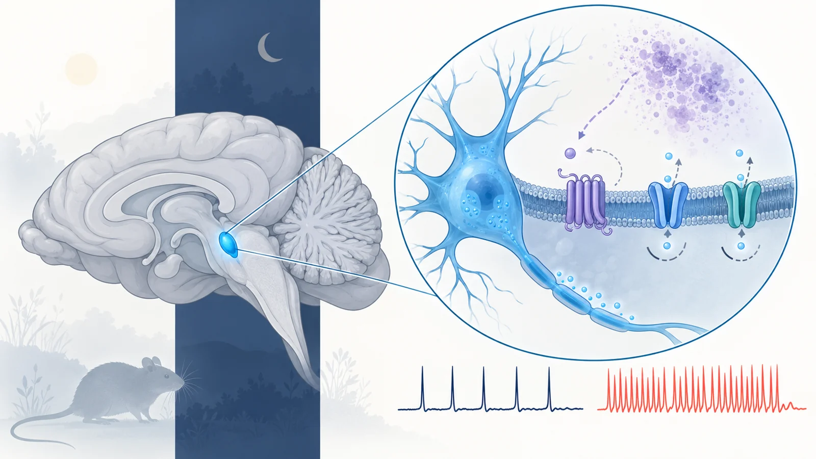
The researchers connected this behavior to the locus coeruleus, a small brainstem region that helps regulate arousal. They then followed the electrical mechanism that could keep this circuit active.
A brainstem arousal circuit loses part of its braking system
Locus coeruleus neurons help promote wakefulness, and their activity also contributes to transitions between brain states. In slices from the young 5xFAD mice, these neurons fired at higher tonic rates during the dark phase than neurons from wild-type mice. More than 65% of the 5xFAD neurons fired above 9 hertz, compared with less than 10% of the wild-type neurons, and the average firing rate was about twice as high.
The timing mattered. During the light phase, the researchers found no genotype difference in tonic locus coeruleus firing. This makes the result different from a circuit that is simply overactive at all times. The abnormality emerged when the mice’s arousal system was expected to be operating most strongly, matching the dark-phase sleep and wake findings.
Potassium channels help impose limits on neuronal firing. After a neuron becomes electrically active, potassium currents help shape the return toward a less excitable state and can restrain further firing. The study focused on low-threshold currents carried in part by Kv4 channels, which contribute to an acute current, and Kv7 channels, which contribute to a more persistent current.
Both forms of low-threshold potassium conductance were impaired in locus coeruleus neurons from 5xFAD mice. The high-threshold currents showed only mild changes. When researchers inhibited Kv7 channels in wild-type neurons, the cells became more excitable. Blocking Kv4 channels also increased firing. These perturbations made the channel findings functionally relevant: reducing either part of this electrical brake could reproduce features of the overactive state.
The impairment did not appear to result simply from having fewer channels. Measurements of major Kv4 and Kv7 subunits found no significant genotype differences in messenger RNA or total protein. The early defect therefore looked more like altered regulation or channel function than a straightforward loss of channel abundance.
Adrenergic signaling normally helps keep firing in check
The proposed regulator was the α2A adrenergic receptor, an inhibitory receptor on locus coeruleus neurons. When norepinephrine release rises during sustained activity, these receptors can act as an auto-inhibitory feedback system, reducing further firing and norepinephrine release. In practical terms, the same circuit that helps generate arousal also contains a mechanism for preventing arousal from running unchecked.
In the 5xFAD neurons, α2A receptor signaling was less effective. Guanfacine, an α2A agonist, still suppressed firing, showing that inhibitory signaling was preserved. But its effect began more slowly and was smaller than in wild-type neurons under conditions when receptor activation was otherwise expected to be low. The receptor’s measured messenger RNA and total protein levels were not significantly different between genotypes, again pointing toward impaired function rather than reduced receptor abundance.
Activating α2A receptors in 5xFAD neurons increased both the acute and persistent low-threshold potassium currents and reduced their hyperexcitability. Clonidine produced a similar effect. Conversely, blocking α2A receptors in wild-type neurons created a hyperexcitable pattern resembling the 5xFAD state.
The findings support a receptor-to-channel pathway in which weakened adrenergic auto-inhibition leaves Kv4- and Kv7-related conductances less able to restrain locus coeruleus firing. They do not establish that this is the only route by which the channels become dysfunctional. Changes to channel modification, interacting proteins, cellular trafficking or the surrounding signaling environment could also contribute.
Amyloid exposure reproduced the electrical problem in a controlled test
The study used two different kinds of amyloid evidence, and they answer different questions. First, measurements showed higher total Aβ42 in the medial brainstem region containing the locus coeruleus than in several other examined regions of two-month-old 5xFAD mice. Amyloid plaques were absent from the locus coeruleus region, and inflammatory markers were minimal at this early stage.
A regional measurement does not show how much amyloid is present inside an individual locus coeruleus neuron, nor does it identify the precise molecular form reaching a particular receptor. It establishes a local amyloid context, not a direct molecular event at the cell surface.
The second experiment was more direct but used a different preparation. The researchers exposed acute slices from wild-type mice to amyloid oligomers. The treated neurons developed higher excitability and tonic firing, along with reductions in both acute and persistent low-threshold potassium conductance. Their electrical state resembled the one observed in young 5xFAD mice.
Guanfacine reversed the amyloid-oligomer-induced hyperexcitability in those wild-type slices. This induction-and-reversal sequence strengthens the case that amyloid-related signaling can engage the α2A–potassium-channel pathway. It still does not prove the exact physical interaction proposed between amyloid oligomers and the receptor, or show that every step of the pathway is required in living animals.
Two drugs improved sleep through related but distinct routes
The researchers next asked whether changing the pathway could alter the animals’ sleep and wake behavior. They infused guanfacine directly into both sides of the locus coeruleus shortly before the dark phase. Compared with vehicle treatment, the mice spent less time awake and more time in NREM sleep during the dark phase. The number of transitions between vigilance states showed a trend toward improvement, but the result should not be presented as complete normalization of sleep organization.
They also tested retigabine, a drug that opens Kv7 channels. When delivered locally to the locus coeruleus, retigabine reduced hyperarousal and restored NREM sleep measures in the 5xFAD mice. This intervention acted closer to the electrical braking mechanism itself, while guanfacine acted through the impaired adrenergic regulator that normally helps control it.
The local administration matters because it ties the observed sleep changes to manipulation of the locus coeruleus area. It gives the anatomical interpretation more weight than a whole-body treatment alone, although it does not mean that all consequences of the drug were confined to one precisely defined cell population.
A separate experiment used a single intraperitoneal injection of retigabine. This systemic treatment increased NREM sleep and the number of wake-to-NREM transitions during the dark phase. It did not change NREM delta power, a measure of the slow electrical activity associated with that sleep state. The result therefore points to improvements in the amount and organization of selected sleep states, not a universal correction of every sleep feature. Because the drug circulated through the body, the systemic experiment also cannot isolate the locus coeruleus as its exclusive site of action.
The result is a mechanism in mice, not yet a human treatment claim
The study’s strength is the way its measurements connect across levels. The mice showed a phase-specific sleep and wake phenotype. Their locus coeruleus neurons showed phase-specific hyperactivity. Electrophysiology identified impaired low-threshold potassium conductance, while pharmacological tests linked Kv4, Kv7 and α2A receptor function to neuronal excitability. Amyloid-oligomer exposure reproduced key electrical changes in wild-type slices, and targeted interventions improved selected outcomes in the living mouse model.
The scope remains specific. The experiments mainly involved young male 5xFAD mice and wild-type littermates, with most work performed at two to three months of age. A mouse model with early amyloid pathology does not establish that the same receptor–channel pathway causes sleep disruption in people with Alzheimer’s disease. Nor do better NREM measures or more frequent transitions establish prevention of cognitive decline, prevention of Alzheimer’s disease or sustained treatment efficacy.
Sleep is made of separable dimensions: time awake, time in NREM or REM sleep, transitions between states and the electrical features within those states can change independently. The intervention results should therefore be read as evidence that selected parts of the sleep–wake phenotype were modifiable, not as proof that the entire architecture was restored.
Human studies will need to determine whether α2A receptor regulation and Kv4/Kv7 function are altered in comparable early disease settings. They will also need to establish whether changing this pathway can produce durable sleep benefits, preserve necessary waking function and remain safe for people. Those translation questions are central because an arousal circuit must be restrained at the right times, not simply suppressed.
What this could change
A treatment that safely reduces excessive arousal while preserving flexible waking could eventually help people with early Alzheimer’s-related sleep disruption spend more time in sustained sleep and move more normally between sleep and wake states. The mouse findings provide a possible route to that capability: repair the adrenergic feedback that regulates locus coeruleus neurons, strengthen the potassium currents that limit firing, or combine both approaches.
That prospect depends on the pathway also operating in humans and on benefits that last beyond a single experimental dose. If those conditions are met, sleep-focused interventions aimed at the biology of excessive arousal could become more precisely matched to an individual’s early disease mechanism, rather than treating disrupted sleep as an isolated symptom.
Key Sources
- Primary source: Impaired adrenergic regulation of Kv channels underlies LC hyperactivity and early-onset sleep disruption in AD-like amyloidogenic mice.
DOI or Journal Page · Peer-Reviewed Paper · 2026-02-01