
Evidence date · 2024-05-06 · Research note 1 of 2
1 2
See current Track status →
Focal cortical dysplasia type II is a developmental brain malformation in which some cortical cells become unusually large and disorganized. In children, this form of dysplasia can be linked to epilepsy that does not respond adequately to anti-seizure medication. Surgery may therefore be needed to remove the brain region generating seizures.
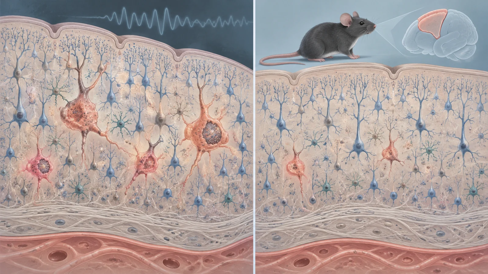
A study published on May 6, 2024, explored a different way to intervene: identify the abnormal cells themselves and remove them selectively. The researchers found senescence-like features in pathological cells from human FCDII tissue, then tested dasatinib and quercetin—drugs used together as a senolytic treatment—in a mouse model carrying a focal, mosaic mTOR-related mutation. In those mice, the treatment reduced measurements of abnormal cells and seizure frequency.
The result is an experimental bridge, not a treatment established for children. Human tissue supplied the biological clues; the intervention and seizure outcome were demonstrated in mice.
Why a small group of cells matters
FCDII is associated with post-zygotic, or somatic, mutations that arise during development rather than being present in every cell from conception. The mutations can affect genes in the PI3K-AKT-mTOR pathway, producing a mosaic pattern: a minority of mTOR-hyperactive cells sits among cells that appear more typical.
The affected cells include dysmorphic neurons, which are enlarged and accumulate neurofilaments, and, in some FCDII cases, balloon cells with both glial and neuronal features. The size of the lesion depends partly on when the mutation arises during development. In some cases, only a small fraction of the resected tissue carries the variant; in larger malformations, the abnormal population can be more extensive.
That arrangement creates a possible treatment opportunity. A therapy would not necessarily need to alter every cell in the brain. It might instead need to affect a pathogenic population that is concentrated within the epileptogenic region. But finding such a population is only the beginning. Researchers would still need to show that the cells are linked to the electrical disorder, that they can be targeted safely, and that removing them changes seizures without damaging useful tissue.
Electrical activity followed the abnormal cells
To examine the human connection, the researchers studied surgically removed cortical tissue from children and adolescents with drug-resistant epilepsy. The larger histological investigation included 22 FCDII specimens and 13 epilepsy-control specimens. A separate ex vivo electrophysiology experiment used tissue from two patients whose slices could be kept oxygenated for recordings after surgery.
The team aligned electrical recordings with whole-tissue images showing where dysmorphic neurons were located. Areas with spontaneous epileptiform discharges contained a higher density of cytomegalic, pS6-positive neurons, a marker pattern consistent with mTOR activity. The measured density was 53.6 dysmorphic neurons per square millimeter in regions showing spontaneous epileptiform events, compared with 11.7 per square millimeter in areas showing only multi-unit activity. Electrically silent regions did not contain the same dysmorphic neurons.
The recordings also helped separate cell types. Balloon cells were found in a different area near the gray-white matter boundary, and that region did not show the spontaneous discharges or multi-unit activity recorded near the dysmorphic neurons. This spatial pattern supports an association between the abnormal neurons and local epileptiform activity. It does not show that every dysmorphic neuron generates a seizure, or that the cells alone explain the entire disease process.
The cells carried a senescence-like signature
The next question was what kind of state these pathological cells occupied. Cellular senescence is an altered cell state marked by changes in gene activity, signaling, lysosomal behavior and interactions with surrounding tissue. It is often discussed in the context of aging, but the researchers were looking for a similar biological signature in pediatric brain tissue—not claiming that the children’s brains had simply aged prematurely.
In the FCDII specimens, the enlarged dysmorphic neurons and balloon cells showed several senescence-associated features. These included activity detected by senescence-associated beta-galactosidase staining, expression of p53 and p16, increased lysosomal content, changes involving the nuclear envelope, and production of a senescence-associated secretory phenotype, or SASP. The markers were concentrated in the pathological cells rather than in neighboring normal-sized cells or the non-mTOR-related control tissues.
The genetic results strengthened the case that the marked cells were part of the disease-associated population. When cells positive for both senescence-associated beta-galactosidase and mTOR activity were isolated from two FCDII tissues, the mutation signal was enriched compared with bulk tissue. In the MTOR case, the variant allele frequency was 42% in the selected cells versus 5.5% in the bulk sample. In the PIK3CA case, it was 41% versus 21.2%.
Those findings supplied a possible target, even though neurons are not dividing cells in the way that many classic senescent-cell models are. The authors emphasize that the biology of senescence in post-mitotic neurons remains poorly understood. The evidence identifies a recognizable state in the pathological cells; it does not by itself establish that neuronal senescence causes seizures.
A senolytic test in a mosaic mouse brain
For the treatment experiment, the researchers used a focal mouse model carrying the Mtor S2215F variant in a mosaic pattern. The model was designed to reproduce important features of FCDII, including abnormal migration, mTOR-hyperactive enlarged neurons and recurrent spontaneous electro-clinical seizures.
The researchers selected a combination of dasatinib and quercetin, known in this experimental context as the DQ senolytic cocktail. The drugs were given by mouth for nine days to mice that had already developed epilepsy. The aim was to reduce the population of cells with the senescence-like phenotype, rather than simply suppress mTOR activity in every affected cell.
The tissue measurements changed after treatment. One month after dosing ended, the density of pS6-positive neurons in the targeted area was nearly 70% lower in DQ-treated mice than in vehicle-treated mice. Senescence-associated beta-galactosidase-positive enlarged cells fell by about 80%. In the remaining dysmorphic cells, pS6 levels were not reduced, suggesting that the treatment did not simply turn down mTOR activity inside every cell that remained. It was associated with a lower number of the marked cells.
These percentages describe cell measurements, not the size of the seizure reduction. The researchers evaluated seizure frequency separately using video EEG recordings.
The seizure response extended beyond the dosing period
Seizure frequency varied among the mosaic mice before treatment. To reduce the effect of that animal-to-animal variation, the researchers used a longitudinal design in which each mouse served as its own comparison. The animals were recorded before and after a vehicle course, then received the DQ combination and were recorded again at the end of treatment and afterward.
The vehicle period did not produce a meaningful change in seizure frequency. After DQ treatment, the researchers observed a statistically significant reduction in the average number of daily seizures. The authors also reported no seizure recurrence during the observation period extending up to one month after the drugs were stopped.
That timing matters because it is consistent with a treatment that lowers the number of seizure-associated cells rather than one that only changes electrical activity while the drug is present. It remains a bounded mouse observation, however. It does not establish lifelong seizure freedom, and it cannot establish how long any benefit would last in a person.
The researchers also tested whether DQ acted as a general anti-seizure treatment in a separate chemically induced seizure model. In that experiment, the drug combination did not significantly change seizure severity compared with vehicle, and senescence-associated beta-galactosidase activity was not detected in the animals used. This response pattern supports a context-dependent interpretation: the treatment appeared more relevant when a senescence-like pathological cell population was present. It does not prove that senescent-cell clearance is the only mechanism involved.
What translation would have to solve
The first unresolved issue is causal biology. The human experiments connect dysmorphic-neuron density with local electrical activity and show a shared cellular signature, but they do not demonstrate the sequence by which the cells produce seizures. The researchers also do not yet know precisely how DQ clears brain cells or what compensatory changes may follow their removal.
Safety is especially important in a developing brain. Senescence-related states can have beneficial roles during development and in normal tissue. A drug that reaches the brain could affect cells outside the pathological population, including cells whose removal would not be helpful. The proposed value of selective clearance therefore depends on finding a sufficiently specific difference between mutated, seizure-associated cells and the surrounding tissue.
The timing of the cell state is another open question. In the human samples, the developmental point at which senescence-like features emerge could not be determined. In the mouse models, the researchers observed these features after birth, around 8 to 10 weeks of age. That animal timeline cannot be transferred directly to children with FCDII.
The experimental scale also calls for proportion. The treatment groups were small, and no statistical sample-size calculation was performed in advance. The researchers report blinded assessment and randomized allocation for the in vivo experiments, which strengthen the design, while the small groups still limit how broadly the findings can be interpreted. The work supports further investigation of a targeted strategy; it does not establish efficacy across all FCDII genotypes, other mTOR-related disorders or epilepsy generally.
What targeted cell removal could mean for epilepsy surgery
For children whose seizures arise from a focal FCDII lesion, the long-term possibility is more selective control of the cells driving the disorder. If future work can identify those cells reliably, remove them without harming surrounding developing brain tissue, and produce durable seizure control in people, a drug-based approach might reduce the need to remove a larger epileptogenic region surgically.
That possibility remains conditional on several difficult steps, including safe delivery, precise biological targeting and sustained clinical benefit. The May 2024 mouse result supplies a reason to pursue those steps: a short course of senolytic treatment was followed by fewer marked pathological cells and fewer seizures in a focal mosaic model. It is an experimental foundation for a potential alternative or complement to surgery, not an established replacement for surgery in children.
Key Sources
- Primary source: Targeting pathological cells with senolytic drugs reduces seizures in neurodevelopmental mTOR-related epilepsy.
DOI or Journal Page · Peer-Reviewed Paper · 2024-05-06