
Evidence date · 2026-03-20 · Research note 3 of 3
1 2 3
See current Track status →
Since the previous research note · Earlier expectation not tested
Track continuity
Since the previous research note: How do immune cells change an aging stem-cell culture?
Previous expectation: More consistent expanded stromal-cell preparations could eventually improve the usable supply of material for regenerative treatment if reproducibility, cell separation, safety and patient benefit were established.
What this study adds: The March 20, 2026 study describes a different route, using temporary factor delivery in umbilical cord-derived stromal cells to establish expandable clones with distinct molecular profiles and some measured functional differences. It does not test the earlier lymphocyte coculture method or its predicted therapeutic reliability.
What changed in this study
Mesenchymal stromal cells are tissue-supporting cells studied for their potential roles in repair and immune regulation. They are also difficult to manufacture consistently. As cultures are expanded, the cells can enter replicative senescence: telomeres shorten, growth slows, metabolism changes and inflammatory signals associated with the senescence-associated secretory phenotype can rise. That matters because cell-based treatments may require very large numbers of cells, yet producing more cells is useful only if the resulting material still behaves like the intended starting population.
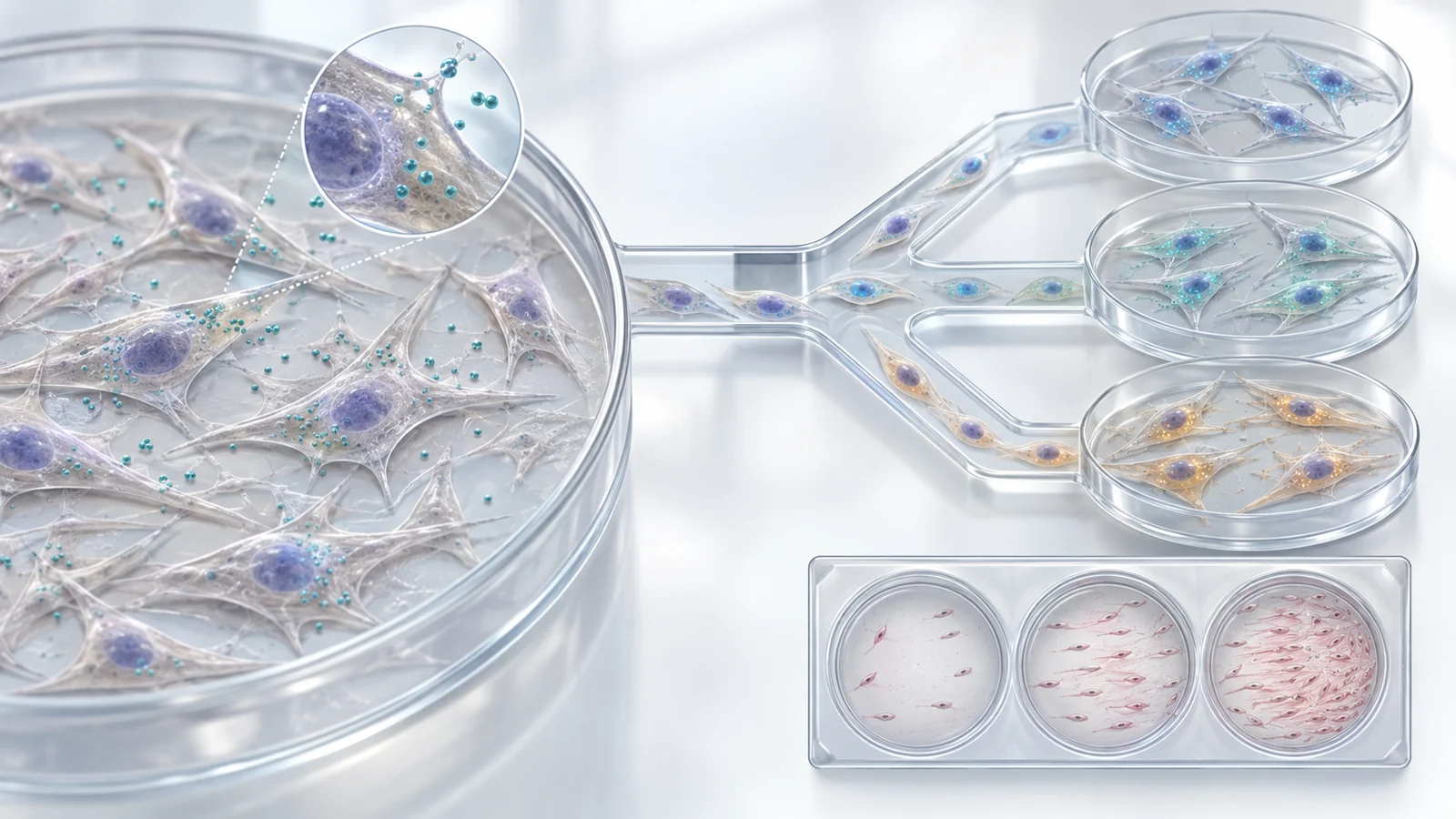
A research article published on March 20, 2026, tested a way to extend the proliferative life of human umbilical cord-derived mesenchymal stromal cells while preserving their identity. The researchers used a temporary, non-integrating delivery system and then separated the resulting cultures into individual lines. The outcome was not one uniform rejuvenated cell product. It was a collection of cell lines with shared stromal features and different molecular and functional profiles.
Why longer growth is only the starting point
The manufacturing problem is easy to state but harder to solve. A culture that stops dividing too soon limits the supply of material. A culture that keeps dividing but changes its behavior may be equally difficult to use. For a regenerative-cell bank, growth capacity needs to sit alongside identity, genomic stability, differentiation competence and reproducible function.
The study therefore examined several properties at once. It measured how long the cells continued proliferating, whether telomeres became longer, whether chromosomes remained apparently stable, whether senescence-associated expression programs were reset, and whether the cells retained the ability to differentiate under the tested conditions. It also looked at individual clones rather than treating every surviving cell as interchangeable.
That choice is important. A bulk culture can conceal meaningful subpopulations. Two samples may both reach a desired cell count while secreting different factors or responding differently in a functional assay. The researchers’ goal was consequently not just to delay culture exhaustion, but to see whether partial reprogramming could produce a scalable library whose members could be characterized separately.
A temporary reset followed by vector removal
The intervention used a temperature-sensitive Sendai virus vector carrying three factors named hTERT, BMI1 and SV40T. The vector operates in the cell’s cytoplasm rather than integrating its genetic material into the cell’s genome. Its encoded factors remain active while the vector is present, and the vector can be passed to daughter cells during cell division.
In the protocol, human umbilical cord-derived cells were exposed to the vector and later shifted to a higher temperature to promote clearance. The cultures were then maintained at normal culture temperature, and fluorescence associated with the vector was used to help identify cells that had lost the vector. Some populations were sorted as bulk SeV-negative cells. Other lines were established by limiting dilution so that individual clonal populations could be studied.
Timing affected the result. Removing the vector between days 20 and 60 preserved subsequent proliferation, while later removal reduced growth. The researchers selected removal at day 20 for the downstream work. This makes the reset a timed intervention rather than an open-ended attempt to keep reprogramming factors active indefinitely.
The supplied assays describe successful removal in different ways at different stages. Vector-encoded expression was undetectable in removed bulk cells by RNA sequencing, while minimal levels were detected in some removed clonal-cell analyses; reverse-transcription qPCR of the clonal cells did not detect Sendai virus at early, middle or late culture stages. The practical point is that clearance was tested directly and was part of the process, not assumed from the temperature shift alone.
What the study meant by rejuvenation
Here, rejuvenation was an operational description of a particular cellular state. The authors looked for extended replicative lifespan and telomere elongation, continued mesenchymal identity without joint expression of the core pluripotency triad, and regenerative or immunomodulatory expression patterns. This is different from saying that the cells became pluripotent or that a person’s biological age had been reversed.
The growth results supported a longer-lived culture under the conditions tested. Vector-cleared clones proliferated for more than 90 days. One clone reached up to 10¹⁶ cells, while three other clones reached approximately 10¹⁰ cells. Separately, the study reported growth beyond 100 days in infected cultures, a result that should not be confused with the post-clearance duration observed for the clones.
Telomeres became significantly longer after vector exposure in bulk cultures, including measurements taken around days 20 and 50, and remained longer than controls after clearance in the day 34–35 removed cultures. Among four analyzed clones, three had longer telomeres than the uninfected control. Telomere shortening was observed after vector removal, so the result was not a claim that length would remain fixed throughout expansion.
The cells also retained mesenchymal markers and the ability to differentiate into neurogenic, adipogenic, osteogenic at an early stage, and chondrogenic lineages under the tested conditions. The propensities were not identical: some lines showed stronger osteogenic, adipogenic or chondrogenic tendencies. These results support preserved differentiation competence in the tested assays, not mature function of every possible derivative or proof that all properties would persist in a clinical manufacturing process.
Expression of the pluripotency-associated factors was examined as another boundary on the state being created. The authors reported no joint expression of the core triad OCT4, SOX2 and NANOG, although low-level expression of an individual member could occur. They used this pattern, together with retained mesenchymal identity, to characterize lineage-preserving partial reprogramming rather than conversion into an induced pluripotent stem-cell population.
A library can contain different biological options
The most consequential finding was not simply that the cells grew longer. The cleared clones displayed different transcriptional profiles, including variation in inflammatory and regenerative genes such as IL6, IDO1, PTGER2 and HGF. Some lines had more pro-inflammatory tendencies, while others showed profiles associated with regenerative or immunosuppressive activity.
Hepatocyte growth factor, or HGF, provided a bridge between a molecular difference and one measured behavior. The researchers found higher HGF secretion in some clones and tested conditioned media in an endothelial-cell migration assay. The migration assay reported stained-cell area percentages of 47% for the high-HGF clone and 13% for the lower-HGF clone. This linked a clone-level expression pattern to protein secretion and to a specific in vitro response.
The result remains narrower than a claim about tissue repair. Migration of endothelial cells in a dish does not demonstrate restored tissue function, the formation of new functional blood vessels in an organism or patient benefit. HGF may be an important contributor in this assay, but the authors also note that other secreted factors could be involved.
The study proposed that differently profiled clones might eventually be matched to distinct research or therapeutic tasks. It also suggested combinations of lines for neuroregeneration-oriented or cartilage-repair-associated programs based on their expression patterns. Those combinations are hypotheses about diversification, not demonstrated synergy. Establishing a useful match would require protein-level confirmation, context-specific functional assays and testing beyond the culture system.
Expansion can change the profile
A cell bank is not characterized once and then understood forever. Continued passage changed expression patterns in this study. HGF tended to decline with prolonged culture, while IL6 gradually increased. A favorable early profile could therefore shift as the cells are expanded, even when the culture continues to grow.
This creates a separate production question from the initial rejuvenation question. Researchers would need to know not only which clone grows efficiently, but also how its secreted factors, inflammatory signals, differentiation behavior and other relevant properties change across the intended expansion window. Growth capacity, stable function and mature differentiation are related requirements, but they are not substitutes for one another.
The differentiation results reinforce that point. Lineage-marker expression did not always predict the measured differentiation outcome. Correlations involving markers such as MAP2, SOX2 and ENO2 were not sufficient to establish regenerative competence. Functional testing remained necessary under the conditions relevant to a proposed use.
A larger library could make this characterization task more useful, because it would offer more candidates to compare. It could also make the task more demanding. The study itself describes its clone collection as a beginning and calls for analysis of substantially more clones, together with direct testing of whether distinct profiles and growth capacities correspond to appropriate functions.
What the safety checks can and cannot tell us
The use of SV40T makes the safety question especially visible. The authors identify it as an oncogene, yet report that transient exposure through the non-integrating, temperature-sensitive vector did not produce a detectable increase in chromosomal instability under the study conditions. Karyotyping found no recurrent numerical or structural abnormalities in the examined infected bulk cells through 100 days or in the removed clones.
The paper also acknowledges numerical deviations of 45 or 47 chromosomes in roughly 5–11% of analyzed cells. Similar low-frequency deviations appeared in parental cells and exposed cells, and the reported abnormalities did not involve recurrent or specific chromosomes. The authors interpret these observations as consistent with baseline variation or technical effects in metaphase spreading rather than an added instability signal from the transient SV40T exposure.
Selected oncogenesis-related transcripts, including MYC, CCNE1, AKT1, BIRC5 and MDM2, did not show persistent overexpression after vector removal, although transient expression varied among cell lines. These are bounded laboratory observations. They do not establish the absence of tumor risk, prove therapeutic safety or replace broader safety testing such as defined genome-wide and in vivo assessments.
The same caution applies to vector clearance. Fluorescence sorting, RNA measurements and reverse-transcription qPCR provide evidence about the assays and samples examined, but they are not a universal guarantee that every future production lot will behave identically. A clinically relevant process would need its own validated release and safety criteria, along with evidence that the relevant properties remain stable during manufacture.
What expandable cell banks could mean for regenerative medicine
The practical promise is a renewable bank of distinct, characterized stromal-cell lines rather than a single population assumed to be uniform. If the growth, identity, functional and safety properties can be reproduced across larger clone collections and maintained through defined expansion, researchers and cell-therapy developers could have a more dependable way to supply material for different repair or immune-regulation tasks.
Such a bank could also make matching more deliberate. A line selected for a particular secreted-factor profile might be investigated for one context, while another line with a different differentiation or inflammatory profile could be evaluated elsewhere. The benefit would remain conditional: favorable behavior in culture must survive production-scale expansion, standardized manufacturing, mature functional testing and rigorous safety evaluation before it can translate into reliable patient benefit.
The March 20, 2026 study provides an early laboratory basis for that capability. Its contribution is the combination of temporary factor delivery, vector removal, clonal separation and side-by-side profiling. Whether those libraries become useful regenerative products depends on the less visible work that follows: proving that a promising clone remains a promising clone as it is made, tested and used.
Key Sources
- Primary source: Rejuvenation of mesenchymal stromal cells via partial reprogramming enables scalable generation of transcriptionally diverse MSC libraries.
DOI or Journal Page · Peer-Reviewed Paper · 2026-03-20