
Evidence date · 2025-11-25 · Research note 2 of 3
1 2 3
See current Track status →
Since the previous research note · Partly supported
Track continuity
Since the previous research note: Can we grow enough useful cells for regenerative treatment?
Previous expectation: Growing enough cells while preserving their useful functions could make future regenerative treatments more dependable, provided production and patient benefit are validated.
What this study adds: Lymphocyte coculture improved functional measurements in expanded human amniotic membrane MSCs, and pretreated cells improved specified outcomes in a mouse colitis model. This supports part of the biological premise without testing manufacturing consistency across settings or benefit in patients.
What changed in this study
A culture can contain many cells and still become less useful over time. Mesenchymal stem cells, or MSCs, may lose some of their ability to divide, form colonies, respond to signals, and develop into different cell types as they are expanded in the laboratory. The problem is not simply producing enough cells. It is maintaining a population with the functions that make those cells useful.
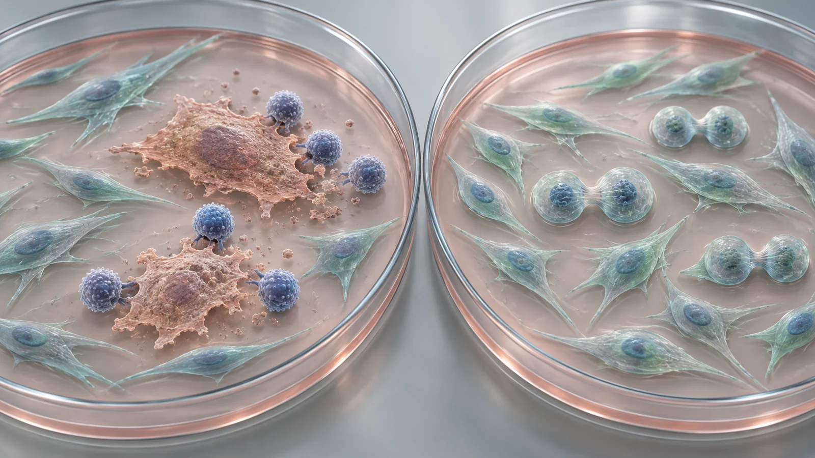
A study published on November 25, 2025, tested whether human peripheral blood lymphocytes could help rebalance such a culture. The researchers worked with human amniotic membrane MSCs, abbreviated hAMSCs, and compared cells at passage 4 with cells at passage 10. A passage is a round of subculture in the laboratory, not a measure of the donor’s age. The later-passage population had undergone more expansion and showed more features associated with cellular senescence, meaning a state in which cells have largely stopped dividing.
The proposed intervention was coculture with peripheral blood lymphocytes, or PBLs. These are immune cells obtained from blood. The central question was not whether every old cell could be made young again. It was whether the immune cells could change the balance of the population by removing some damaged cells while helping other cells continue to grow.
The experiment changed the mix inside a growing culture
Long-term expansion can make an MSC culture increasingly heterogeneous. Some cells retain stronger growth and differentiation properties, while others accumulate stress-related changes. Senescent cells can also release factors that influence nearby cells, making the culture less consistent. For a cell-based therapy, that mixture matters because a high cell count does not guarantee a high-quality product.
The researchers treated passage 10 hAMSCs with PBLs and then assessed several features rather than relying on one marker. PBL-treated cultures formed more colonies, a measurement of how effectively cells can generate new groups of cells. The treated passage 10 cells also showed higher levels of the stemness-associated factors Nanog and Sox2, approaching the levels seen in the earlier-passage comparison. Their relative telomere length, cell-cycle activity, and proportion of EdU-positive cells also improved in the reported experiments.
The functional changes were not identical across all lineages. PBL treatment enhanced osteogenic differentiation, related to bone-forming potential, and chondrogenic differentiation, related to cartilage-forming potential. Its effect on adipogenic differentiation, related to fat-cell formation, was small. This pattern is more informative than a general claim that the cells became universally younger: particular properties changed by different amounts.
The overall picture is therefore population-level rejuvenation. The culture contained fewer cells with strong senescence features and a larger or more active share of cells with useful growth and stemness characteristics. That does not show that every senescent cell individually returned to an earlier state. The authors also could not completely exclude the possibility that some aged cells entered renewed replication, because senescent cells are not all biologically identical.
Contact mattered more than adding signals alone
PBLs release cytokines and other molecules, so the researchers tested whether soluble inflammatory signals by themselves could explain the change. They examined several cytokines whose levels increased in the coculture, including CXCL1, CXCL8, CCL2, IL-33, IL-1α, and IL-1β. These treatments did not reproduce the full reduction in the senescence phenotype seen with PBLs. IL-1α and IL-1β did increase cell numbers and promoted division, but they were less effective than the lymphocyte treatment in the broader comparison.
The physical arrangement of the cells also mattered. Direct coculture produced stronger anti-senescence effects than a transwell setup that kept the two cell populations separated while allowing soluble factors to pass between them. This supports a role for cell-to-cell contact or contact-dependent signaling. It does not eliminate every possible contribution from soluble molecules, nor does it establish exactly how the immune cells recognize their targets.
The comparison helps locate the intervention more precisely. The result was not simply the consequence of adding a generic inflammatory mixture to an old culture. PBLs, acting as cells in direct contact with hAMSCs, produced a wider response that included both selective loss of some cells and stronger expansion of others.
Two mechanisms helped reshape the population
The clearance side of the response involved apoptosis, a form of programmed cell death. In the passage 10 cultures treated with PBLs, more cells carried both a senescence-associated β-galactosidase marker and cleaved caspase 3, a marker associated with apoptosis. The overlap increased by approximately threefold compared with untreated passage 10 cultures. This supports the interpretation that cells with senescence features were preferentially entering programmed cell death rather than simply disappearing from the measurement.
Molecular findings pointed toward a p53-dependent mitochondrial pathway. PBL treatment increased SUMO-1 and strengthened the association between p53 and SUMO-1. SUMOylation means attaching a small protein tag to another protein, which can alter where that protein acts and how it behaves. The study also found changes consistent with p53 moving toward mitochondria, increased cytochrome C, and a shift in apoptosis-related proteins. Together, these results support the proposed route from PBL contact to p53 regulation and mitochondrial apoptosis. They do not by themselves prove that this pathway alone accounts for every cleared cell.
The growth side of the response involved Serpinb2 and NF-κB signaling. Serpinb2 increased in the treated passage 10 cells, and reducing Serpinb2 prevented the PBL treatment from lowering the senescence marker p21, increasing proliferation, or raising Sox2 and Nanog levels. NF-κB also moved into the nucleus in the coculture, where it was associated with the proliferation marker Ki67. Blocking NF-κB activation reduced Ki67 expression, while Serpinb2 knockdown weakened the related signaling change.
These perturbation experiments give the growth explanation more support than a simple correlation would. They indicate that Serpinb2 is needed for much of the proliferative response observed in this system and that NF-κB signaling participates in it. The culture was being remodeled through two linked but distinct processes: apoptosis reduced part of the aged population, while signaling helped relatively younger cells divide and retain useful characteristics.
The treated cells were tested in an inflamed mouse colon
The study also asked whether the altered cells behaved differently in a disease model, rather than only in laboratory assays. The researchers used chemically induced colitis in mice and randomly assigned six animals to each of four groups: normal mice, mice receiving the colitis-inducing treatment, mice receiving passage 10 hAMSCs, and mice receiving passage 10 hAMSCs pretreated with PBLs.
The disease was induced with DSS in the drinking water. The hAMSCs were injected through the tail vein on the first and fourth days of DSS administration, before the severe symptoms reported from day five. The experiment therefore tested early intervention during disease induction, not treatment begun after severe colitis was established. The researchers then assessed body-weight change, disease activity, colon length, tissue appearance, inflammatory cytokines, and intestinal barrier-related proteins.
Compared with mice given untreated passage 10 hAMSCs, the PBL-pretreated cells improved the reported weight, disease-activity, and colon-length outcomes. The treated-cell group also showed stronger changes in the reported histological, inflammatory, and barrier measurements, including IL-1β, IL-6, Occludin, Claudin-1, and Claudin-2. In this experiment, the coculture intervention was associated with a measurable functional difference in an animal model.
That result extends the finding beyond cell-cycle markers, but it remains a small preclinical experiment. The mouse model does not establish that the same preparation would work in people, and the specified improvement in colitis does not show that the proposed rejuvenation mechanism alone caused the therapeutic effect.
Safety checks left questions about clinical use
Introducing immune cells into a manufacturing process creates a practical identity question. The PBLs were allogeneic to the MSCs, meaning they came from a different genetic background and could be more immunogenic. In the coculture, the lymphocytes outnumbered the MSCs by about 100 to 1. The researchers washed the PBLs away after 72 hours and examined the remaining cell population.
Before subculture, 2.6% of the collected cells were positive for the MHC class II markers HLA-DR, DP, and DQ. The authors interpreted this initial signal as potentially reflecting lymphocytes that had not been completely removed, given the large difference in starting cell numbers. After the MSCs were subcultured, MHC class II expression was negative in the tested population. The reported G-banding results also indicated that the coculture did not change the hAMSC karyotype.
The study included a tumorigenicity test with five nude mice per group, comparing passage 10 hAMSCs, PBL-pretreated passage 10 hAMSCs, and A549 cells. The MSC-injected animals were followed for three months, and the authors reported no tumor masses after transplantation of the PBL-pretreated cells. This is a limited safety observation in mice, not proof of long-term safety in patients; a future manufacturing process would still require reliable removal and characterization of the added immune cells.
The surviving senescent fraction is another unresolved part of the process. PBL treatment reduced the aged-cell population but did not eliminate every cell with senescence features. Given the heterogeneity of senescent cells, the authors could not exclude the possibility that PBL treatment stimulated renewed replication in some aged cells. That possibility matters when a culture is intended for expansion, even though it does not negate the improvements measured in this experiment.
What better cell cultures could mean for future therapies
A method that shifts an expanded MSC culture toward more dividing, colony-forming, and functionally responsive cells could eventually help manufacturers produce a larger usable supply of therapeutic material rather than merely a larger number of cells. For patients, the potential benefit would be indirect at first: more consistent cell preparation could become part of a more dependable route to regenerative treatment.
That possibility is conditional. The coculture approach would need reproducible performance across donors, passages, laboratories, and MSC sources; controlled removal of the added lymphocytes; adequate safety validation; and evidence that the resulting cells benefit patients. As of the study’s November 2025 publication, the evidence shows a promising way to reshape human amniotic membrane MSC cultures and improve outcomes in a mouse colitis model. It does not yet show that the approach can deliver a safe, effective cell therapy in people.
Key Sources
- Primary source: Rejuvenation of mesenchymal stem cells by human peripheral blood lymphocytes.
DOI or Journal Page · Peer-Reviewed Paper · 2025-11-25