
Research Signal – Source date 2025-09-19
Managing high blood pressure, diabetes, cholesterol, and smoking is central to reducing vascular harm to the brain. Yet established cognitive decline can persist because the injury may already involve more than the risk factors that helped start it. Damaged small vessels can reduce blood flow, weaken the blood–brain barrier, sustain inflammation, and injure the white matter that carries signals between brain regions.
A review published on 19 September 2025, Vascular Dementia: From Pathophysiology to Therapeutic Frontiers, brings these processes together and surveys possible treatment directions. It describes a field without an approved disease-modifying therapy specifically for vascular dementia (VaD). The review’s proposed treatments remain largely investigational, so their biological rationale should not be mistaken for demonstrated benefit in patients.
Vascular dementia can begin with different kinds of injury
Dementia associated with vascular disease does not arise through one uniform route. After a stroke, cognitive impairment may follow a single infarct in a region important for cognition, or build as multiple infarcts damage neural circuits over time. The review defines post-stroke dementia as dementia developing within a year after a clinical stroke. Stroke severity and location, recurrence, and a person’s cognitive status before the stroke all affect the risk.
Small-vessel disease can cause a more gradual pattern. In subcortical ischemic vascular dementia, damage to small arteries and arterioles is linked to chronic reductions in blood flow to deep brain areas. MRI may show white matter hyperintensities, small deep infarcts called lacunes, microbleeds, and enlarged spaces around vessels. The associated difficulties can include slower processing, problems with executive tasks, and changes in gait; memory may be relatively preserved early on.
The categories can overlap with Alzheimer’s disease. The review describes mixed vascular and neurodegenerative pathology as common in older people with dementia. Vascular lesions can add to cognitive decline and lower the threshold at which Alzheimer’s pathology becomes disabling. In the other direction, amyloid in vessel walls can compromise vascular integrity. A vascular-versus-degenerative label may therefore fail to capture the combination of processes affecting a person.
How small-vessel injury can disrupt brain networks
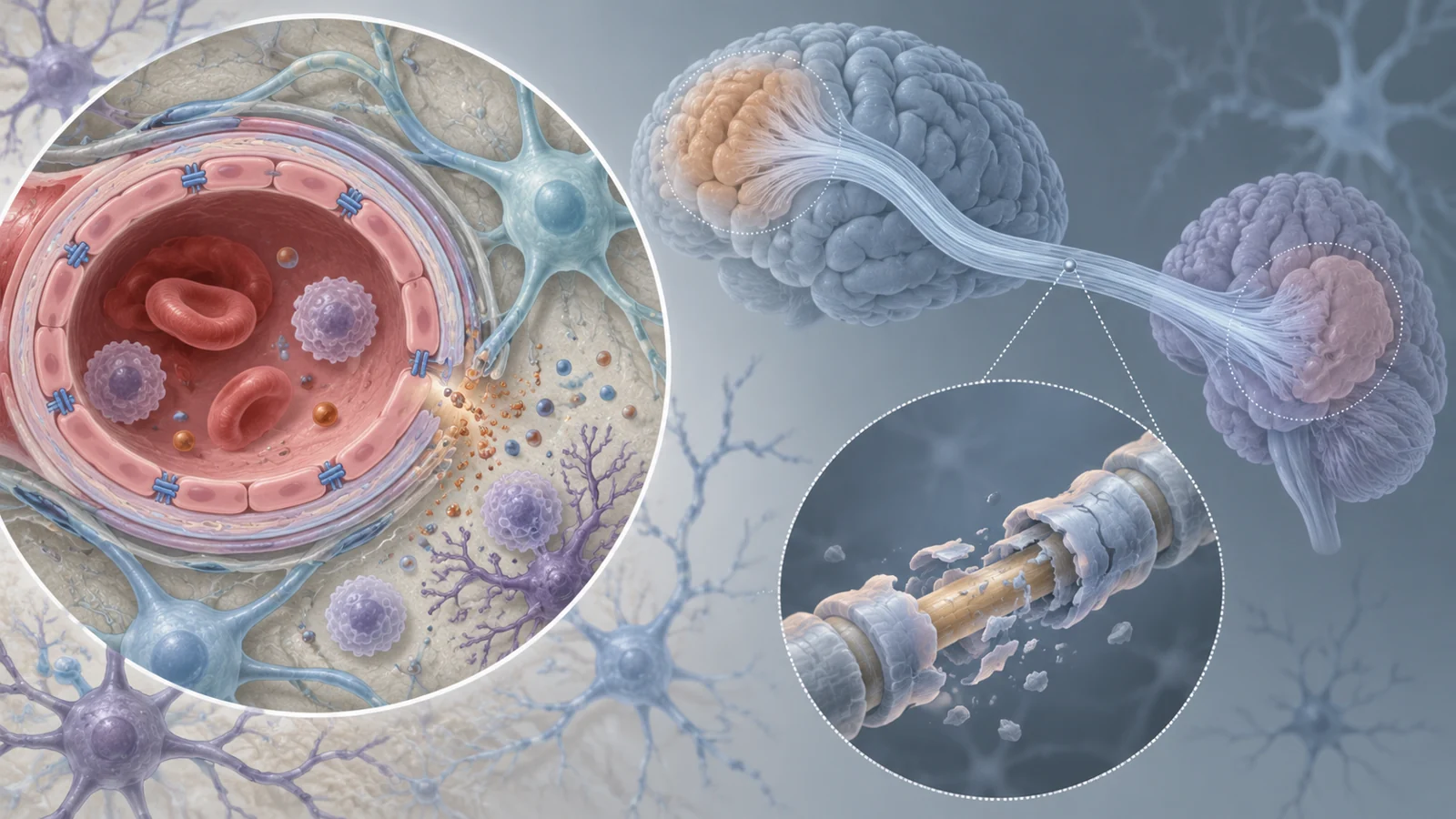
Healthy brain function depends on a neurovascular unit: endothelial cells lining vessels work alongside pericytes, astrocytes, and neurons. In the review’s account, chronic low blood flow and vascular risks can push endothelial cells away from a stable state. They produce less of the vessel-relaxing molecule nitric oxide and more of the constrictor endothelin-1, while also displaying signals that encourage inflammation and the movement of immune cells into brain tissue. These changes can further compromise circulation.
The vessel lining also helps form the blood–brain barrier, which normally regulates what passes from the bloodstream into the brain. In vascular injury, connections between endothelial cells can break down. The resulting leakage lets blood proteins, including albumin and fibrinogen, enter brain tissue. The review links albumin to astrocyte activation and inflammation, and fibrinogen deposits to impaired repair of myelin, the insulating material around nerve fibers.
Inflammation and oxidative stress can harm oligodendrocytes, the cells that maintain myelin. The environment may also interfere with precursor cells that could replace lost oligodendrocytes. As myelin and the underlying axons are damaged, communication between distant brain areas becomes less reliable. Injury to these connections offers one explanation for slowed processing and executive difficulties in small-vessel disease: the problem can involve the routes linking brain regions, not just a single site.
The review also discusses impaired glymphatic clearance, a brain-wide process associated with removing waste, among the mechanisms that may contribute to injury. Reduced arterial pulsation and loss of pericytes are proposed as contributors to early dysfunction. These interacting pathways help explain why controlling vascular risks remains essential but may not, by itself, repair established damage to barriers, cells, myelin, and neural connections.
The proposed sequence is a research model, not a personal timetable
The review organizes disease processes into a possible progression. Before clear symptoms, endothelial dysfunction and small increases in barrier permeability may appear alongside early signs associated with glymphatic impairment. With emerging cognitive symptoms, barrier leakage, oxidative stress, and a more persistent inflammatory response may coincide with stress to oligodendrocytes. In established disease, the review describes stronger inflammatory activity, more extensive myelin and axonal injury, and damage affecting wider brain networks.
This sequence gives researchers a reason to ask whether a treatment aimed at vessels or the barrier would be most useful at a different stage from one intended to limit inflammation or support myelin repair. It is not a clock for diagnosing an individual. VaD includes different patterns of vascular injury, and the course can depend on the extent of that injury and a person’s susceptibility.
Scans and fluid markers answer different questions
MRI provides visible evidence of vascular injury. White matter hyperintensities can reflect chronic damage and gliosis; lacunes and microbleeds add other clues to small-vessel disease. The amount and progression of white matter changes can relate to cognitive decline. More advanced imaging, including diffusion tensor imaging and arterial spin labeling, may help investigate early white matter changes and blood flow when standard scans leave uncertainty.
Fluid markers can add information that a scan cannot supply on its own. Neurofilament light chain in cerebrospinal fluid or blood can indicate axonal damage. Inflammatory and endothelial markers may offer clues about active processes. Alzheimer’s-related amyloid and tau measurements can help identify possible mixed pathology. The review notes that combining MRI features can increase diagnostic confidence, while the performance of imaging measures varies across patient groups, definitions, and scanning protocols. No single marker described in the review provides a validated rule for assigning a person to one of the proposed therapies.
Proposed treatments still need clinical validation
Risk-factor management remains current practice. The review emphasizes controlling high blood pressure, diabetes, high cholesterol, and smoking, particularly for prevention. It also notes that these measures have limited ability to reverse or halt cognitive decline once it is established. That gap motivates efforts to target the tissue injury and biological pathways that may continue after vascular damage begins.
Among the approaches it surveys are phosphodiesterase inhibitors intended to support cerebral perfusion; inhibitors of the NLRP3 inflammasome, an inflammatory platform; senolytics aimed at senescent cells; and agents intended to promote remyelination. Strategies to strengthen the blood–brain barrier are another proposed direction. These ideas address different parts of a connected process, but the review does not demonstrate that any of them slows cognitive decline in patients. It describes most mechanism-directed approaches as investigational and points to limited Phase III evidence.
Moving from a plausible target to useful care requires clinical studies to show that an intervention improves outcomes. It will also matter whether a therapy reaches the process driving injury in a particular group of patients. Biomarkers could eventually help characterize that process, but the reviewed markers have not yet established a treatment-selection rule. Evidence that a pathway changes is not by itself proof that people think or function better as a result.
What targeted care could mean for people with vascular dementia
If reliable markers can identify the dominant injury process and clinical studies show that matching treatments improve outcomes, care might pair vascular risk control with therapies aimed at the brain damage that persists. Addressing the relevant process could give some people a better chance of preserving thinking for longer. That possibility depends on validated ways to select treatment and evidence of benefit in patients; the 2025 review describes a research direction, not a proven treatment strategy.
Key Sources
- Primary source: Vascular Dementia: From Pathophysiology to Therapeutic Frontiers.
DOI or Journal Page · Review or Meta-Analysis · 2025-09-19
Keep exploring