
Research Signal – Source date 2021-01-21
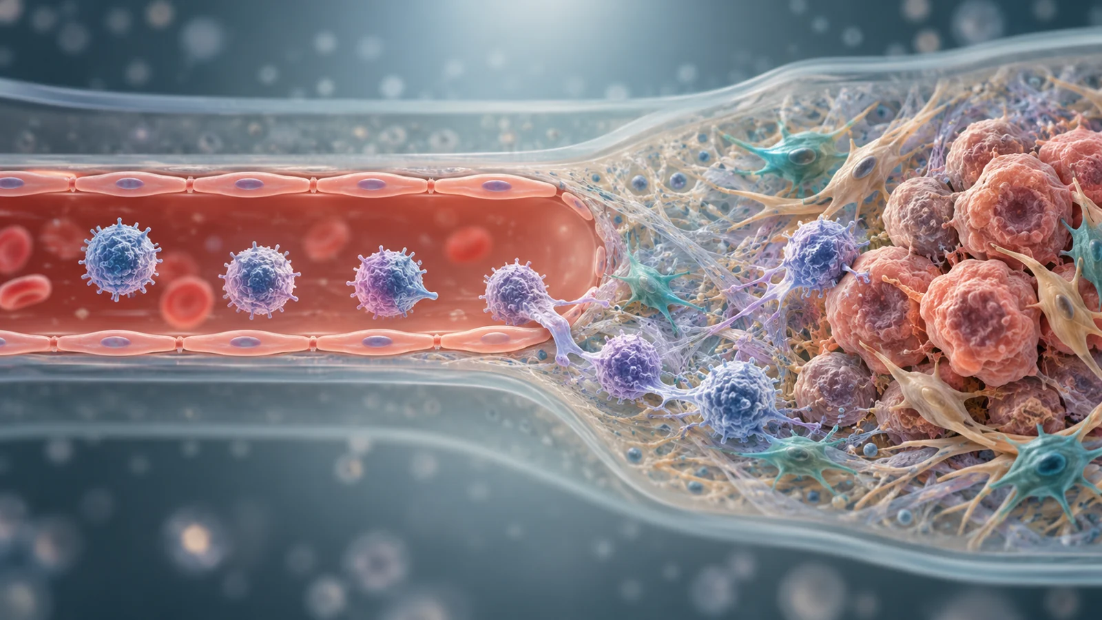
A tumor-on-a-chip is a small laboratory model designed to bring parts of a tumor environment together. To study how immunotherapy might work, researchers need to consider more than what happens after an immune cell reaches a tumor. The cell’s route toward the tumor, including its passage through a vessel lining, is part of the interaction too.
A 2021 review describes microfluidic devices as a way to model these connections and study cancer–immune system interactions. It discusses platforms for solid tumors and blood cancers, and emphasizes the vessel-like barrier that immune cells cross on their way toward a tumor site.
The route into a tumor is part of the interaction
The tumor microenvironment is not just a cluster of cancer cells. It includes tumor cells, stromal cells—which support or surround the tumor—and immune cells moving toward the site. Their relationships can change as cells arrive and conditions in the environment shift.
In the review’s account, immune cells reach the tumor through an endothelial barrier: a lining associated with blood vessels. Including this boundary in a model gives researchers a way to study immune-cell trafficking, or movement toward and across the barrier, as part of cancer–immune crosstalk. A model focused only on tumor cells would leave that part of the process out.
The review places these models in the context of immunotherapies, treatments meant to help the immune system fight cancer. It names immune checkpoint inhibitors and adoptive cell-based products as examples. The review does not show that a chip can predict whether either treatment works for a patient.
Microfluidics can bring several parts into one model
Microfluidic devices offer a way to construct models with multiple interacting parts of the tumor environment. Rather than treating the tumor as an isolated target, researchers can use these platforms to investigate how tumor and supporting cells relate to immune cells and a vascular component.
The review covers approaches for solid tumors and blood cancers. Those settings are not interchangeable, and the abstract does not establish that one model reproduces them with equal fidelity. Its broader point is that microfluidic platforms can help researchers study cancer–immune interactions while accounting for more than one component of the environment.
Imaging can make changing responses measurable
A model is more useful when researchers can observe how cells respond as the environment changes. The review highlights image processing and related algorithms for real-time monitoring and quantitative evaluation of cellular responses. In practical terms, images can be analyzed over time to track cell behavior and turn visual observations into measurements.
That focus connects the model’s moving parts to a readout: researchers can examine cellular responses as modeled conditions change, rather than relying only on a single snapshot. The review’s abstract describes this as an important part of evaluating these platforms, but it does not provide comparative performance figures for particular devices or establish that their measurements predict an individual patient’s response.
How tumor chips could change preclinical immunotherapy tests
If later studies validate the chip readouts against outcomes beyond the model, these platforms could help researchers detect a problem with immune-cell access before taking a candidate immunotherapy further. That would give them a chance to investigate a failure a simpler tumor-cell test might miss. The review does not establish that chip results predict clinical benefit.
Key Sources
- Primary source: Tumor-on-a-chip platforms to study cancer-immune system crosstalk in the era of immunotherapy.
DOI or Journal Page · Review or Meta-Analysis · 2021-01-21
Keep exploring