
Research Signal – Source date 2026-03-01
Sleep problems are common in Alzheimer’s disease, and they involve more than spending fewer hours asleep. Sleep can become fragmented, deep sleep can diminish, and the rhythms that organize activity across the sleeping brain can change. Those rhythms matter because the brain uses coordinated activity during sleep to process memories.
A review published on March 1, 2026, brings the thalamus into this picture. This deep brain structure is not a single relay station: it contains many nuclei with different connections and jobs. The authors assemble evidence that some thalamic regions change early in Alzheimer’s and explain how thalamic circuits help time sleep rhythms. The proposed connection is compelling, but research in people has not yet shown that thalamic damage drives the sleep and memory changes of the disease.
A relay with several distinct roles
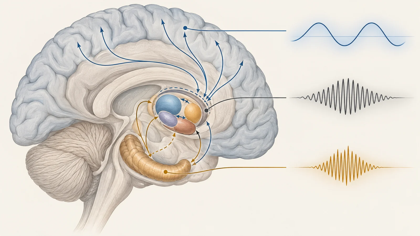
The thalamus sits between the brain’s cortex and other regions, exchanging information with both. Its nuclei differ in their connections: some relay sensory information, while higher-order and limbic nuclei participate in communication among cortical and subcortical regions. Limbic nuclei connect with areas involved in memory and behavior, including the hippocampus and prefrontal cortex.
A thin structure called the thalamic reticular nucleus, or TRN, surrounds much of the thalamus. Its inhibitory neurons regulate the activity of thalamic nuclei and help shape communication between the thalamus and cortex across wakefulness and sleep. The TRN’s role in rhythm generation makes it especially relevant to sleep; other nuclei contribute to communication with memory-related regions.
This division of labor matters. A change in one nucleus may affect a different circuit or function from a change elsewhere in the thalamus. Describing “thalamic damage” as if it were one uniform event would miss that complexity.
Early changes appear uneven across the thalamus
The review brings together findings from people with inherited and sporadic forms of Alzheimer’s, as well as people with mild cognitive impairment. In some presymptomatic carriers of a PSEN1 mutation, studies found reduced thalamic volume or white matter changes before measurable hippocampal volume loss. Other work reported medial thalamic changes alongside Alzheimer’s-related pathology. In sporadic disease, thalamic atrophy has been observed by mild stages, though its timing relative to changes in other brain regions varies across studies.
The affected areas also differ. Findings include changes in anterior and mediodorsal regions, the pulvinar, and nuclei near the midline, among others. Postmortem work described neuronal loss concentrated in nuclei connected with the limbic system. The review also discusses reported atrophy and reduced activity in the TRN.
Communication between the thalamus and other brain regions can change as well. Studies in mild cognitive impairment and Alzheimer’s have found altered functional connectivity, including reduced connections between higher-order thalamic nuclei and cortical areas. One study reported increased connectivity between the left and right thalamus in mild cognitive impairment, a possible compensatory pattern. The evidence is not yet a complete account of how these connections change over time, and results differ by group and method.
Together, the findings make early thalamic involvement plausible in some people, not a universal sequence in which the thalamus changes first. They also leave open whether these structural and connectivity changes contribute to sleep disruption, result from broader disease processes, or reflect both.
Sleep depends on timing across circuits
During non-REM sleep, the cortex produces slow waves. The thalamus helps set their pace and synchronize them across cortical areas, and it is a major generator of delta waves. Different nuclei contribute differently: specific thalamic nuclei pace slow oscillations in sensory cortex, while midline nuclei help synchronize slow activity across regions.
Sleep spindles are brief bursts of rhythmic activity seen mainly in the cortex during non-REM sleep. They begin with rhythmic firing in inhibitory TRN neurons, which shapes activity in thalamic cells. The resulting bursts travel to the cortex and can propagate toward the hippocampus. Spindles are associated with synaptic plasticity and memory consolidation; in studies, stronger spindle activity after learning has been linked with better retention.
These rhythms are useful to understand as a coordinated sequence rather than as separate measures of sleep quantity. Slow waves, spindles, and brief hippocampal ripple events need to align in time for communication among the hippocampus and cortex. In particular, ripples may transfer memory-related information toward the cortex when spindles coincide with the active phase of slow waves.
Alzheimer’s studies find disrupted coordination, not a settled cause
The review describes sleep changes in Alzheimer’s that include less REM and non-REM sleep, reduced time in the deepest stages, and greater fragmentation. It also summarizes altered sleep oscillations in people and animal models. In people with Alzheimer’s, reduced coupling between slow oscillations and spindles has been associated with weaker overnight memory consolidation.
Animal findings add detail. In one mouse model, sleep quantity was preserved while the timing coordination of oscillations was impaired and correlated with spatial memory deficits. Another model showed reduced coupling between hippocampal ripples and prefrontal spindles, even though local ripple structure was preserved.
These observations connect altered rhythm timing with memory measures, but they do not establish that thalamic dysfunction causes cognitive decline in people with Alzheimer’s. The review identifies gaps in the disease-specific evidence: a direct causal link between thalamic dysfunction and slow-wave changes has not been established, and the role of thalamic changes in disrupted ripple–spindle coordination remains open. The circuit explanation provides a direction for research, rather than a demonstrated chain from thalamic damage to sleep disruption to memory loss.
Mouse interventions point to possible targets
Some experiments have tested whether changing thalamic activity can affect sleep or memory in Alzheimer’s models. Chemogenetically activating inhibitory neurons in the TRN increased non-REM sleep and reduced fragmentation in one mouse model; the study also reported lower amyloid plaque burden in the neocortex and hippocampus. Deep brain stimulation of the nucleus reuniens restored thalamus–hippocampus connectivity and improved working memory in APP/PS1 mice. Stimulation of intralaminar nuclei also improved cognitive measures and synaptic plasticity in a rodent model.
These results show that selected thalamic circuits can be manipulated in animals with measurable effects. They do not establish a safe or effective direct thalamic treatment for people with Alzheimer’s. Reaching specific nuclei is difficult because the thalamus is deep and anatomically complex.
Other human findings discussed in the review concern a different approach. Non-invasive stimulation during sleep has enhanced slow oscillations and their coordination with spindles, with memory benefits reported in healthy people and people with mild cognitive impairment. Those findings concern sleep-rhythm stimulation; they do not demonstrate direct thalamic treatment of Alzheimer’s disease.
What sleep-rhythm treatment could change in Alzheimer’s
For people with Alzheimer’s, a treatment that restores coordinated sleep activity might help preserve the overnight processing of memories and could potentially reduce disease-related harm. That possibility depends on identifying which nuclei and rhythms matter, finding a safe way to modulate them in people, and showing a clinical benefit. The animal results offer a reason to investigate the thalamus; they are an early step toward that goal, not evidence that such a treatment is ready.
Key Sources
- Primary source: Implication of the thalamus in sleep alterations observed in Alzheimer's disease.
DOI or Journal Page · Review or Meta-Analysis · 2026-03-01
Keep exploring