
Evidence date · 2024-12-28 · Research note 2 of 3
1 2 3
See current Track status →
Since the previous research note · Partly supported
Track continuity
Since the previous research note: How paired nighttime brainwave and muscle measurements revealed different neurological group patterns
Previous expectation: Repeated home sleep measurements might eventually help clinicians follow neurological profiles if the patterns generalize and track meaningful disease changes.
What this study adds: The December 2024 study expanded the measurements into four-category profiling and assessed repeat assignments in small longitudinal subsets, providing partial support for persistence of profiles. It did not establish independent generalization or validate profile changes as measures of disease course.
What changed in this study
A sleep recording can contain more than a bedtime summary. In a December 2024 study, researchers used several overnight measurements and age to assign people to broad neurological profiles, then asked whether those assignments resembled their existing clinical diagnoses and whether they remained similar when testing was repeated.
Those are related questions, but they are not the same one. A profile may agree with a diagnosis at one assessment yet change later. It may also remain stable while still being an imperfect representation of the underlying condition. The study offers an exploratory look at how far sleep measurements might support neurological assessment, while leaving the clinical value of the profiles to be tested independently.
Turning an overnight recording into a profile
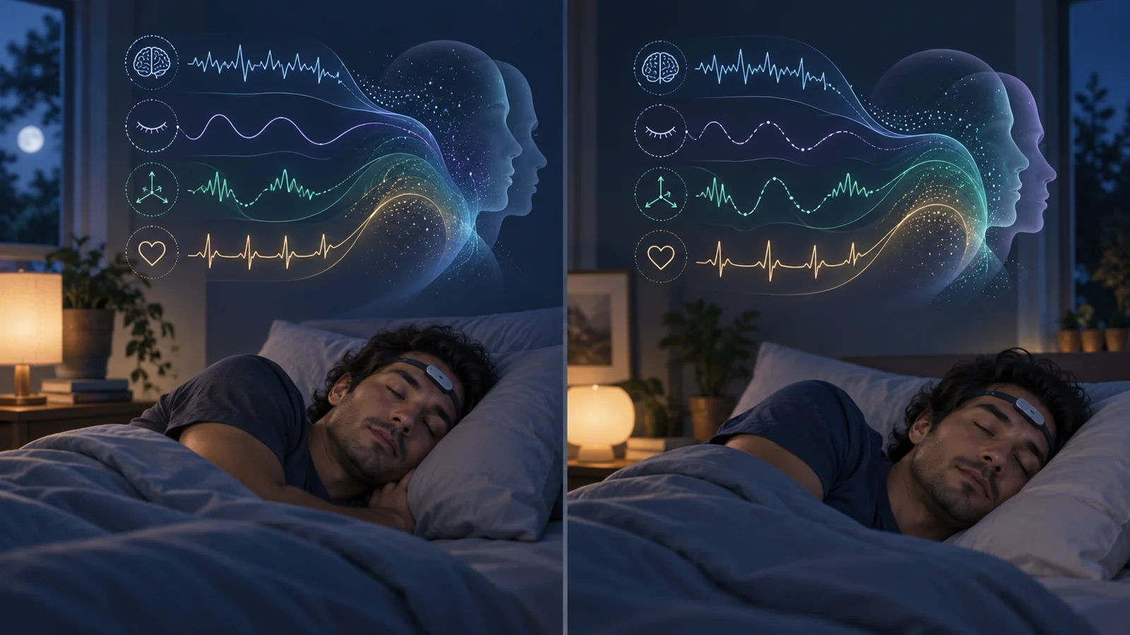
The recordings came from a forehead-based sleep system that measured frontopolar electrical brain activity, eye movements, pulse, head movement and position, and snoring. Most participants were studied at home; 18 people with isolated REM sleep behavior disorder were recorded during laboratory polysomnography.
The researchers extracted nine sleep biomarkers and combined them with age. These included REM duration, sleep efficiency, time spent asleep on the back, pulse-related autonomic activation, sleep-spindle duration, non-REM hypertonia, and measures of non-REM brain activity. The classifier used these derived measures rather than every signal the device recorded; snoring was not among the nine listed biomarkers.
A four-category machine-learning model was trained with groups representing Alzheimer’s disease dementia, Lewy body dementia, isolated REM sleep behavior disorder, and controls. The category trained on isolated REM sleep behavior disorder participants was used as a profile for possible prodromal synucleinopathy. REM sleep without normal muscle relaxation was not one of the classifier’s inputs, so the label was derived from the category structure rather than from directly feeding that feature into the model.
When more than one night was available, the biomarker values were weighted across nights according to the amount of sleep represented by each recording. The model then produced probabilities across its categories, which were translated into labels such as Probably Normal, Likely Normal, or a probable neurological profile. These labels described the model’s output; they were not grades of evidence and did not replace the participants’ clinical diagnoses.
What counted as agreement
The study evaluated 381 recordings, including the records used in model development and additional recordings from people with Alzheimer’s disease, Parkinson’s disease without dementia, mild cognitive impairment, isolated REM sleep behavior disorder, and controls. That design allowed the researchers to compare the distribution of profiles across several clinically different groups, but it was not a test in a wholly independent population.
Agreement had to be defined before the percentages could be interpreted. A control participant was counted as aligned when the model produced a Probably Normal or Likely Normal profile. For a participant with a diagnosed disorder, a disease-compatible probable or likely profile counted as agreement. A Likely Normal result with indications pointing toward the person’s diagnosis was treated as a trend toward agreement, rather than the same as a straightforward disease-compatible classification. Conversely, a Probably Normal profile in someone with a diagnosed disorder, or a profile pointing to an incompatible disorder, was treated as an overt disagreement.
The model’s clearest group-level patterns appeared in Alzheimer’s disease, Lewy body dementia, and controls. The paper reports disease-compatible or normal classifications in more than three quarters of those groups, depending on the category and how borderline indications were counted. Among controls, most profiles were Probably Normal or Likely Normal, although a small group received probable neurological profiles. In Alzheimer’s disease, most participants received a Likely or Probable neurological profile. Lewy body dementia showed a similar broad alignment, with some profiles marked normal or pointing toward Alzheimer’s disease.
The pattern was less uniform in the other groups. Just over half of the isolated REM sleep behavior disorder participants received a Likely or Probable neurological profile, while others were classified as probably normal or assigned a profile that did not fit the clinical category. Parkinson’s disease and mild cognitive impairment also showed mixtures of compatible, normal, and conflicting assignments. The authors interpreted many of the apparent disagreements as potentially reflecting differences in disease severity or in how much abnormal sleep physiology was present, rather than as a clean separation between true and false classifications.
A repeat result answers a different question
The longitudinal analysis was small and varied in timing. Forty-six controls were retested between eight and 58 months after baseline. The dementia follow-up subsets included ten people with Lewy body dementia and nine with Alzheimer’s disease, with retests at six and 12 months; not everyone contributed a retest at both time points.
The most useful way to read these results is to separate identical labels from similar profiles. Seventy-four percent of controls received exactly the same profile at baseline and retest. The broader abstract measure, which included the same or a similar profile, was 98% for controls. For Lewy body dementia, 70% had the identical profile, while 88% had the same or a similar assignment. For Alzheimer’s disease, the corresponding figures were 79% and 86%.
A stable label did not necessarily mean a correct diagnosis. One participant with Alzheimer’s disease was characterized as Probably Normal both at baseline and again 12 months later. Other participants moved between probable, likely, mixed, and normal-with-indications categories. A person with Lewy body dementia shifted from a probable Alzheimer’s profile to a probable mixed profile and then to a probable Lewy body profile. These changes show that the classifier could preserve a broad pattern while still shifting in its more specific interpretation.
Repeat testing therefore addressed persistence of the model’s output, not whether a changing output tracked the course of disease. The follow-up records were repeated observations from small subsets, not independent patients added to the original evaluation. Nor did the study show that changing a profile would predict a symptom change, treatment response, or future clinical event.
Why the profiles can overlap
The model was trying to distinguish conditions that can share sleep-related features. The discussion identifies reduced spindle duration and increased non-REM slowing as abnormalities that may occur in both Alzheimer’s disease and Lewy body dementia. When those shared features dominate, the probabilities may not cleanly separate the categories, producing a mixed label or a classification that appears to point to the other disorder.
The individual measurements also have clinical context. The authors note that a high autonomic activation index, abnormal spindle duration, and extended time sleeping supine could prompt consideration of untreated obstructive sleep apnea. Medications can influence the measurements as well: some antidepressants were associated in the authors’ prior work with increased non-REM hypertonia and suppression of REM sleep, while benzodiazepines may affect sleep efficiency and produce activity resembling sleep spindles. These examples do not turn the classifier into a diagnostic checklist. They show why a clinician would need to inspect the contributing signals and the person’s circumstances rather than treat a profile label as self-explanatory.
The study’s reference categories also leave important biological ambiguity. Participants were assigned clinical diagnoses without pathological confirmation or the deeper imaging and fluid-biomarker characterization that could help distinguish Alzheimer’s, Lewy body, or mixed pathology. A profile that agrees with a clinical category can therefore indicate concordance with that category without proving the protein changes presumed to underlie it.
The evidence is promising but still preliminary
The study’s performance estimates need to be read in light of how the evaluation was assembled. The 381-recording set included training records, and the same Lewy body records were used for model development and validation. The authors acknowledge that this could inflate the apparent performance in that subgroup. Independent cross-validation and larger cohorts would be needed to determine how well the profiles generalize beyond the data used here.
The control group also relied on the Mini-Mental State Examination rather than more detailed neuropsychological testing. Some control participants received profiles suggestive of hidden disease, and several were later excluded from the control test-retest analysis after their cognitive scores declined. Two participants who initially had high scores and profiles with Alzheimer’s indications later had lower scores and probable Alzheimer’s profiles; another older participant had a probable Alzheimer’s profile at both time points alongside a later lower score.
Those observations are important for understanding the study’s uncertainty, but they do not establish that the sleep model predicted future dementia. These were observations in only a few people, who were excluded from the control retest analysis after their cognitive scores declined. The paper’s central result is therefore about exploratory classification and repeat assignment, not early diagnosis, prevention, or improved care.
Possible mixed Alzheimer’s and Lewy body pathology adds another limit. Treating the groups as if each person belonged to one exclusive category made the classification task workable, but real clinical cases may not fit that separation. The study itself calls for more detailed clinical assessments, imaging, fluid biomarkers, and longitudinal sleep measurements before the classifier could be considered a dependable clinical tool.
What repeat sleep profiles could add to neurological care
Repeated home recordings could eventually give clinicians an additional physiological view of a person’s neurological condition between conventional assessments. The practical appeal is continuity: a profile could be checked again without requiring every follow-up to repeat a full laboratory sleep study, while the component measurements could help show which parts of the pattern changed or remained similar.
That possibility depends on several links holding together. Larger, independent cohorts would need to confirm that the profiles generalize. Future longitudinal work would need to show that meaningful changes in a profile correspond to meaningful changes in symptoms, cognition, or disease biology. Clinical interpretation would still need to sit alongside established assessments, because a persistent classification can be wrong and a shifted classification can reflect overlapping conditions, medication effects, or other influences on sleep.
As of December 2024, the study supports a narrower conclusion: sleep-based profiling showed enough group agreement and repeat consistency to justify further testing as an adjunctive physiological measure. It did not yet show a validated diagnostic test, a reliable measure of progression, or a demonstrated benefit for patients. The potential value lies in whether repeated, interpretable sleep measurements can add useful information to care once those questions are answered.
Key Sources
- Primary source: Concordance and test-retest consistency of sleep biomarker-based neurodegenerative disorder profiling.
DOI or Journal Page · Peer-Reviewed Paper · 2024-12-28