
Research Signal – Source date 2026-04-01
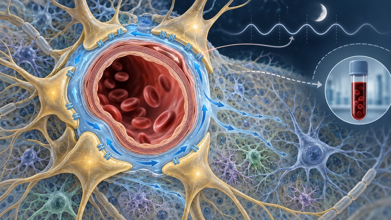
A result described in a 2026 narrative review sounds backwards at first. In a randomized crossover study of 39 adults, morning blood levels of several amyloid and tau species were higher after normal sleep than after a night of sleep deprivation. If amyloid and tau are associated with Alzheimer’s disease, why would more of them appear in the blood after sleep?
The answer may depend on where the proteins are measured and what happened to them overnight. A higher concentration in blood does not automatically mean that more protein accumulated in the brain. It could also reflect material leaving the brain more effectively. The same measurement can therefore carry different meanings depending on production, release, transport, removal, sampling time, and physiological state.
The review, published on April 1, 2026, brings together animal experiments, human biomarker studies, imaging research, and small intervention studies. Its central subject is the glymphatic system, a proposed brain-wide fluid-clearance network that may become especially active during natural sleep. The evidence makes sleep biologically important to Alzheimer’s research while leaving open how much better sleep can do for long-term memory.
The blood result that seems backwards
A morning blood sample captures what has reached the bloodstream at that moment. It does not directly report how much amyloid or tau remains in brain tissue, how much was produced during the night, or how quickly material moved between the brain, cerebrospinal fluid, and blood.
Sleep can affect several of those processes at once. The review discusses human and experimental evidence that sleep loss can increase amyloid concentrations in fluid surrounding the brain, with stable-isotope work pointing toward increased production during wakefulness. Tau also appears sensitive to disrupted sleep. Normal sleep, meanwhile, may improve the movement of some molecules from the brain toward the blood.
Those observations can coexist. Higher morning plasma levels after sleep were interpreted by the review as consistent with more effective overnight brain-to-blood clearance. Lower levels after sleep deprivation could occur alongside greater production or retention in another compartment. A blood result therefore cannot be read as a simple score for either accumulation or removal.
The crossover finding is best understood as evidence about overnight kinetics rather than a direct demonstration that sleep removed plaques or repaired memory-related circuits. Protein concentrations reflect competing processes, and the direction of a short-term change depends on when and where the sample was taken. The surprising result opens the mechanistic question; it does not settle the clinical one.
A fluid network that works with the vessels
The glymphatic model describes cerebrospinal fluid entering spaces around arteries, exchanging with fluid between brain cells, and leaving through pathways associated with veins and the meninges. Arterial pulsation, vessel movement, and pressure differences may help drive this circulation. The model is useful, but it sits within a broader clearance network that also includes meningeal lymphatics, perivascular drainage, diffusion, and other forms of fluid and solute movement.
Astrocytes occupy an important interface between blood vessels and brain tissue. Their endfeet surround vessels, and the water channel aquaporin-4, or AQP4, is concentrated in those perivascular membranes. In animal experiments, disrupting AQP4 reduced exchange between cerebrospinal and interstitial fluid and slowed the clearance of injected substances, including amyloid-beta. The review also describes how age- or disease-related loss of AQP4’s normal perivascular localization could impair transport even when total AQP4 protein remains present.
Sleep may provide conditions that make this exchange more effective. The review describes evidence that natural sleep and some sleep-like states expand interstitial space and increase fluid exchange in mice. Slow-wave sleep is especially relevant, but newer mechanistic work points to a dynamic process involving infraslow norepinephrine oscillations, vessel movement, and pulsatile cerebrospinal-fluid inflow.
Animal experiments add causal detail to this model. In amyloid models, acute and chronic sleep deprivation increased interstitial amyloid and accelerated plaque deposition. In tau-focused models, sleep loss increased interstitial or cerebrospinal-fluid tau and could worsen tau seeding and spread. Manipulating AQP4 produced related changes in transport and Alzheimer-related pathology. These results support a role for sleep-linked clearance biology in animals, without establishing the size or clinical importance of an equivalent effect in people.
What human measurements can actually show
Human studies approach this biology through measurements that answer different questions. DTI-ALPS is an indirect diffusion-MRI proxy designed to capture water-diffusion patterns near perivascular spaces. In Alzheimer-focused cohorts, lower values have been associated with amyloid burden, neurodegeneration, or disease progression. Its practical appeal is that it is less invasive than introducing a tracer and can be combined with other brain measurements.
Its biological meaning is not specific enough to treat it as a direct readout of glymphatic flow. The review describes population studies in which ALPS was strongly influenced by age, vascular burden, and white-matter abnormalities. A lower value may therefore reflect a mixture of tissue structure, vascular health, and fluid-related changes. Associations with Alzheimer-related pathology can also weaken after vascular and white-matter factors are considered.
Tracer-enhanced MRI provides a different approach by following an introduced substance through cerebrospinal-fluid spaces. It is more direct in the narrow sense that researchers can observe tracer movement, but the procedure is invasive and therefore difficult to use for routine assessment or large, repeated studies. A diffusion proxy and an introduced tracer are not interchangeable measurements, and neither one alone resolves the full transport process.
The same limitation applies to short-term biomarker changes. A shift in an imaging proxy or a morning blood value may show that physiology changed, but it does not by itself demonstrate increased glymphatic transport, reduced brain plaques, improved memory, or prevention of Alzheimer’s disease. Stronger human evidence would need to connect a transport-related measurement with a durable clinical outcome.
Sleep is more than being unconscious
The review’s discussion of interventions shifts attention from the simple question of whether someone slept to the physiological state that sleep produced. If clearance depends on slow-wave activity, norepinephrine rhythms, and healthy vascular oscillations, then unconsciousness alone may not reproduce the relevant conditions.
Mouse work described in the review found that zolpidem suppressed norepinephrine oscillations and reduced glymphatic flow, even though it produces a sleep-like state. This is an experimental result in animals. It does not establish an equivalent effect or a particular dementia risk in people taking the drug. Its importance is conceptual: behavioral sleep and clearance-supporting sleep physiology may come apart.
A separate human study used closed-loop acoustic stimulation and enhanced slow-wave activity at the group level, with marked variation between participants. The result shows that an intervention can influence one feature of sleep, but it does not establish that the intervention improved glymphatic transport or protected cognition. The relevant response may depend on sleep continuity, the timing and structure of slow waves, vascular dynamics, and a person’s underlying age-related or disease-related changes.
This broader view also explains why insomnia, sleep apnea, circadian disruption, and fragmented sleep appear in the review’s account of Alzheimer’s biology. Sleep quality may affect protein handling through several routes, but the therapeutic target would need to be physiologically meaningful sleep rather than sedation, duration alone, or a reassuring change in one isolated biomarker.
What better sleep could mean for Alzheimer’s prevention
For older adults and people at risk of Alzheimer’s disease, the potential benefit is concrete: sleep-focused care could eventually help preserve cognition by supporting the brain’s handling of amyloid, tau, and other proteins. That prospect remains conditional. Researchers first need reliable human measures of relevant transport and then evidence that improving it produces durable cognitive benefit or changes the clinical course of disease.
A validated approach could make sleep part of a broader prevention strategy that also considers vascular health, inflammation, protein biology, and individual risk. It could help distinguish an intervention that changes the physiology relevant to brain protein handling from one that changes sleep duration, sedation, or a short-lived blood measurement.
The review does not establish that sleep-based enhancement of clearance prevents Alzheimer’s disease. It shows why the question is worth pursuing and why the answer cannot come from one surprising blood result. Better sleep may help the brain manage potentially harmful proteins, but protecting memory will require a demonstrated chain from sleep physiology to human clearance and then to lasting clinical benefit.
Key Sources
- Primary source: Sleep-Dependent Clearance of Brain Metabolites via the Glymphatic System: Implications for Alzheimer's Pathophysiology.
DOI or Journal Page · Review or Meta-Analysis · 2026-04-01
Keep exploring