
Research Signal – Source date 2021-11-08
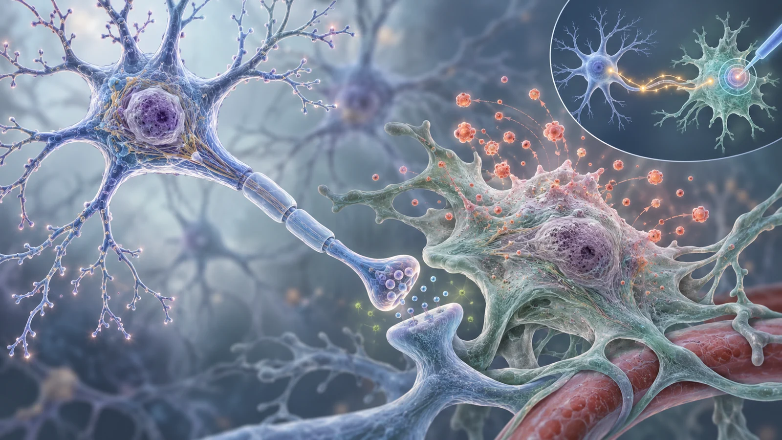
Amyotrophic lateral sclerosis (ALS) destroys the motor neurons that control movement, so a proposal to kill cells in the nervous system sounds counterintuitive. The cells under discussion, however, are not the motor neurons themselves. They are glial cells, including astrocytes and microglia, that normally help maintain the environment around neurons. When some of these support cells enter a persistent stress-associated state called senescence, they may become part of the problem.
A review published on November 8, 2021, proposed senolysis as a possible way to address that problem. Senolytic drugs are designed to remove senescent cells, but the review placed the idea firmly in the research stage. At that publication perspective, senolysis had not yet been evaluated in ALS. The proposal rested on a chain of findings from ALS models, patient-derived cells, human tissue and studies of other neurological diseases, not on demonstrated improvement in ALS movement or survival.
The cells being targeted are not the neurons
Senescence is a non-replicating state that cells can enter after repeated stress or injury. Senescent cells remain metabolically active, resist normal cell death and change their behavior. They also release inflammatory molecules and lose some of the functions that healthy cells perform. In the nervous system, those changes matter because glia are not passive bystanders. They help regulate the chemical and electrical conditions in which neurons operate.
The distinction between a motor neuron and a senescent glial cell explains the apparent contradiction. The proposal is not to remove the nerve cells whose loss causes weakness. It is to remove surrounding cells that may be releasing harmful signals or failing to provide normal support. Whether that could be done safely is the central biological problem, because a pathway that keeps a senescent glial cell alive may also help a stressed motor neuron survive.
Two ways altered glia can strain motor neurons
One route involves inflammation. Senescent cells can develop a senescence-associated secretory phenotype, or SASP, in which inflammatory signals accumulate around them. The review describes astrocytes in ALS as sources of molecules such as TNF-alpha, TGF-beta, IL-6, IFN-gamma and nitric oxide. These inflammatory signals alone do not establish that those cells are senescent. These signals can recruit or activate additional immune responses, creating conditions in which inflammation spreads through neighboring cells rather than remaining confined to the original site.
A second route is the loss of ordinary glial support. Astrocytes help remove excess glutamate from synapses and regulate potassium ions. In the cellular experiments discussed by the review, induced senescence reduced the expression of the glutamate transporters EAAT1 and EAAT2 and the potassium transporter Kir4.1. Senescent astrocytes also showed reduced glutamate uptake, lower mitochondrial activity and more reactive oxygen species. The resulting imbalance could leave more glutamate near motor neurons, increasing excitatory stress. Increased expression of GRINA may add to that concern because the associated NMDA receptor system allows calcium to enter motor neurons.
These are different forms of harm. One involves what altered glia release; the other involves what they no longer clear or regulate. A treatment that removes a harmful cell would need to address both possibilities without removing healthy support cells or damaging neurons that are already vulnerable.
A pattern, not a single diagnostic label
The case for senescence in ALS was assembled from several indicators rather than one definitive test. Senescence-associated beta-galactosidase was elevated in spinal cords from SOD1G93A animal models, and astrocytes derived from those tissues showed related changes. Astrocytes made from induced pluripotent stem cells carrying a C9orf72 mutation also showed higher beta-galactosidase expression and a decline in the proliferation marker Ki-67 over time. Findings involving p21 and other cell-cycle regulators appeared in ALS models, patient-derived astrocytes and post-mortem human material.
The details matter because senescence markers do not all appear at the same point or mean exactly the same thing in every cell. p16 may rise later in senescence, while p21 can be highest during development of the state and then decline. p21 can also indicate DNA damage in some settings. The review therefore treats the combination of markers as more informative than any one result.
Location adds another qualification. In ALS-affected brains, p21 was elevated in both neurons and glia in the frontal association cortex, but not in the motor cortex in the cited finding. That does not describe a uniform population of senescent cells throughout the nervous system. It suggests that cell type, region and disease stage may shape what researchers observe. Patient-derived astrocytes and animal spinal-cord models contribute useful pieces to the picture, but they answer different questions from post-mortem human tissue.
Promising comparisons leave a causal gap
The review draws on three experimentally separate ideas. First, clearing senescent cells improved disease-related measures in models of other neurological disorders. In a tauopathy mouse model, selectively eliminating senescent microglia and astrocytes reduced gliosis, prevented tau phosphorylation and aggregation, and reversed short-term memory loss in that model. Related work in several Alzheimer’s disease models also linked senescent-cell clearance with lower neuroinflammation, less pathology or improved cognitive measures.
Second, interventions that altered inflammation improved outcomes in ALS models. Reducing selected inflammatory signals lessened motor impairment in SOD1G93A mice, while cromolyn sodium reduced microglial activation, delayed symptom onset and was associated with greater motor-neuron survival in the cited experiments. These findings support the idea that inflammatory processes matter in ALS, but they do not show that the interventions worked by clearing senescent cells.
Third, a compound may have more than one relevant action. The review discusses quercetin as a possible senolytic candidate, while also noting its anti-inflammatory and antioxidant effects. If a future ALS experiment showed an improvement after quercetin treatment, the result would not automatically identify senolysis as the cause. The compound’s direct effects on inflammation or oxidative damage would also need to be separated. Evidence from a tauopathy or Alzheimer’s model can make a biological strategy plausible, but it cannot substitute for an ALS test.
The drug target could protect one cell and endanger another
Senolytic design becomes especially difficult when the same survival machinery operates in both harmful glia and vulnerable neurons. The review highlights PI3K and related Akt signaling as an example. These pathways help senescent cells resist apoptosis, making them attractive targets for senolysis. Yet increased PI3K or Akt activity improved viability and motor function in some ALS-related cellular and animal experiments. Blocking the pathway could therefore make senescent glia easier to remove while weakening motor neurons at the same time.
Bcl-2-family proteins offer a different possible route. They help cells resist apoptosis, and the review discusses their inhibition as a way to push senescent cells toward death. Navitoclax cleared senescent cells in several experimental settings and reduced senescence-related pathology in a mouse model of Alzheimer’s disease. But its broader interference with Bcl-2-family proteins was associated with depletion of neutrophils, leukocytes and platelets in humans. More selective compounds directed at Bcl-xL were presented as a possible way to narrow the effect, not as a clinically established ALS solution.
The target-selection problem therefore extends beyond the spinal cord. A candidate would need to distinguish senescent glia from healthy glia, motor neurons and other cells that rely on overlapping survival pathways. The review’s interest in Bcl-2-family inhibition reflects that challenge and the possibility of protecting motor neurons through more than one route. It does not establish that selective glial elimination, neuronal protection or meaningful ALS benefit has been achieved.
What this could change
For people with ALS, the potential payoff is a therapy that acts on damaging surroundings rather than trying to replace the motor neurons already being lost. If researchers can identify senescent glia accurately, clear them without undermining motor-neuron survival and show a meaningful ALS benefit, reducing that source of inflammation and lost support could help preserve movement for longer. The authors also present earlier slowing of progression and possibly longer survival as future aspirations, conditional on solving those selectivity and safety problems. In November 2021, senolysis was a plausible research direction, not an ALS treatment shown to work.
Key Sources
- Primary source: Senolytics: A Novel Strategy for Neuroprotection in ALS?
DOI or Journal Page · Review or Meta-Analysis · 2021-11-08
Keep exploring