
Research Signal – Source date 2022-04-10
A cell can stop dividing and still have a busy biological life. It can consume energy, release signals, and influence neighboring cells. This helps explain why cellular senescence—a state of sustained withdrawal from the cell division cycle—can both protect the body and contribute to disease. Stopping a damaged cell from multiplying can help suppress cancer. Leaving biologically active senescent cells in tissue can have different consequences, depending on what they release and how long they persist.
Senolytics are drugs designed to eliminate senescent cells. But a report that a compound “targets senescence” can describe several different actions. A cell might die, release fewer inflammatory signals, or change its growth behavior. Whether the surrounding tissue works better is a further question.
Published April 10, 2022, Cellular Senescence: Molecular Targets, Biomarkers, and Senolytic Drugs brings together research on how scientists detect these cells, produce them experimentally, and intervene in their biology. It is a review of existing studies. Its useful contribution is to connect those questions, making it easier to understand what a promising drug experiment actually demonstrates.
A living cell with its growth brakes engaged
Senescence and aging are related, but they describe different things. Aging concerns changes across an organism over time. Senescence is a cellular response that can follow repeated division, DNA damage, or other stresses. It also participates in processes such as wound healing and tissue repair.
Senescence is conventionally described as stable, irreversible growth arrest. Yet the review also discusses circumstances in which cells can escape that arrest, particularly in cancer. The term therefore does not mean that a cell is dead, permanently harmless, or simply a miniature version of an old person.
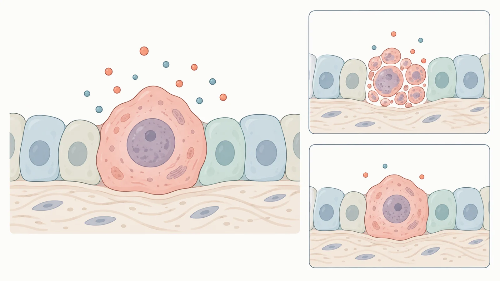
Inside a senescent cell, several systems may change together. Cells often become enlarged and flat. Their waste-processing compartments, called lysosomes, become more active. Survival pathways can make them resistant to apoptosis, the regulated process through which cells die.
Many also develop a senescence-associated secretory phenotype, abbreviated SASP. This means they release a mixture of molecules that can influence inflammation and the behavior of surrounding cells. The mixture and its effects depend on the biological setting. Growth arrest and secretion are connected features, but they are regulated separately enough that a treatment can alter one without reversing the other.
That distinction makes senescence an unusual therapeutic target. The growth brake may be useful, while some consequences of the cell’s continued activity may be harmful.
Finding senescence takes more than one positive stain
The review states that no single marker unequivocally identifies a senescent cell. Researchers instead assemble evidence from several features, interpreted in the context of the tissue and experiment.
One widely used test detects senescence-associated beta-galactosidase, often shortened to SA-β-gal. Its activity is associated with changes in lysosomes. A positive stain can help locate candidate senescent cells, but the enzyme is also active in settings that do not establish senescence, including neurons and developing embryos. Familiarity and convenience do not make it a unique identifying tag.
Other measurements examine the growth brake itself. Researchers can look for proteins such as p16 or p21, which participate in pathways that restrict cell division. They can also check for a reduction in DNA synthesis or the absence of Ki67, a protein associated with actively cycling cells. Enlarged cell shape, altered nuclear structure, and secreted molecules provide additional observations.
These measurements answer related questions rather than duplicating one another. A cell that is not making DNA has stopped progressing through part of the division cycle; that observation alone does not explain why. An enzyme stain reports a biochemical activity. Bringing such observations together makes the interpretation more specific.
The same reasoning matters after treatment. If a senescence-associated marker falls, the cells might have been eliminated—or their marker expression might have changed while they remained alive. Establishing clearance requires evidence about cell survival and abundance. Establishing tissue benefit requires measurements of what that tissue can do. One disappearing stain cannot carry all three conclusions.
Killing a cell and quieting its secretions are different actions
A major senolytic strategy attacks the survival machinery that allows senescent cells to resist death. The review describes compounds that inhibit members of the BCL-2 protein family, which help prevent apoptosis. Disrupting that protection can make susceptible senescent cells die.
Navitoclax is one example. Studies discussed in the review reported depletion of senescent cells in cell experiments and mouse models. Here, the intended action is removal of the cell, with its secretions disappearing as a consequence of its elimination.
Another approach changes secretion while the cell remains present. The review describes experiments in which rapamycin, which inhibits the cellular regulator mTOR, partly suppressed components of the SASP. It also discusses drugs acting through other signaling pathways that reduced secretion in particular experimental settings. A quieter secretory output is a distinct biological result from cell death.
This difference matters when interpreting the broad language of drugs “against senescence.” Reduced inflammatory signaling could change a cell’s effects on its neighbors without reducing the number of senescent cells. Conversely, killing cells need not restore a tissue’s function if other damage persists. The biological action and the desired health outcome need separate evidence.
Restarting division is different again. The review describes experimental ways of bypassing growth arrest, alongside findings that some cancer cells escaping senescence can acquire properties associated with more aggressive tumors. Releasing a damaged cell’s growth brake is therefore not interchangeable with clearing it. Calling both actions rejuvenation would obscure a consequential difference in what happens to the cell.
The experiment determines which question a drug answers
Senescent cells produced by different routes need not respond identically to treatment. The review describes replicative senescence, which researchers can induce by repeatedly growing and dividing cells in culture. In suitable cell types, shortening of telomeres—the protective ends of chromosomes—helps bring that process to a halt.
Radiation-induced senescence starts from a different insult. Radiation damages DNA, and the response depends on dose, delivery schedule, cell identity, and the severity of the damage. Under some conditions cells enter senescence; under others they die. The time allowed after exposure also matters because the senescent state develops over time.
A drug tested against repeatedly divided connective-tissue cells is therefore being examined in a different biological situation from a drug tested against irradiated cancer cells. Neither experiment automatically represents every cell that accumulates during ordinary aging.
Drug examples make that dependence concrete. Studies summarized in the review found senolytic effects of dasatinib in senescent human fat-cell progenitors and of quercetin in cells from the lining of umbilical veins. Results for individual compounds and combinations reflect the cells and conditions tested. They do not create a universal ranking of which drug is best.
Animal experiments add interactions that a dish cannot reproduce. A compound encounters multiple tissues, immune activity, metabolism, and routes of elimination. Those interactions can alter its effects. A mouse model offers a way to study the response of an intact organism, while the way senescence was induced still shapes what the experiment can explain.
Cancer treatment gives senescence two different jobs
Cancer makes the importance of sequence especially clear. A treatment may push tumor cells into senescence, stopping their multiplication. That growth arrest can serve an immediate purpose even though the cells survive.
The difficulty arises if surviving cells persist after treatment. The review discusses therapy-induced senescence as a possible reservoir for later disease recurrence, including circumstances in which cells can escape arrest. This motivates a proposed two-step approach: induce senescence during cancer treatment, then eliminate the remaining senescent cells.
Studies discussed in the review tested navitoclax after chemotherapy-induced senescence in cell and animal models. Other experiments examined radiation-induced senescent cancer cells. The authors also describe animal findings in which treatment reduced metastasis and relapse. These are proof-of-concept findings for a treatment sequence, rather than evidence that the sequence reduces recurrence or mortality in patients.
The two stages have different purposes. The initial intervention restrains tumor growth; the later intervention aims to remove cells that survived in an arrested state. Whether that sequence helps depends on the cancer, the treatment-induced cell state, and the effects of the subsequent drug. Simply counting both stages as “anti-senescence” would miss why senescence is deliberately created before it is targeted.
A useful treatment must work beyond the target cell
A compound can act on the intended biological pathway and still have an unfavorable balance of benefit and harm. Navitoclax and the related compound ABT-737 illustrate the problem. The review reports toxicity toward platelets and neutrophils, blood components involved in clotting and immune defense. Such effects constrain how the compounds might be used clinically.
This is a practical limit on the idea of removing unwanted cells. Survival pathways are not exclusive to the cells researchers want to clear. An intervention must achieve enough of the intended effect without causing unacceptable injury elsewhere.
Delivery systems add another set of questions. The review discusses nanoparticles that can package active compounds and carry coatings intended to influence where they go. Such carriers could change drug exposure in particular tissues. But their distribution, interactions within the body, side effects, and clearance all require investigation. A carrier that performs well in a laboratory experiment has not thereby solved drug delivery in an organism.
As described in the April 2022 review, some candidates, including dasatinib and quercetin and other compounds studied for effects on senescence, had entered human trials for age-related conditions. That places the field at the stage of investigating possible clinical uses. Prior use of a drug for another purpose does not establish the benefit or safety of using it to target senescent cells.
The experimental findings summarized here do not establish human lifespan extension or general rejuvenation. Those claims reach well beyond showing that a compound changes a marker, suppresses secretion, or removes susceptible cells in a model.
What this could change
If selective clearance produces durable tissue benefits with acceptable harm in humans, it could provide a way to preserve function in tissues affected by age-related disease. The causal promise is concrete: removing cells whose persistent activity contributes to dysfunction could reduce that burden on surrounding tissue, allowing it to work better. Treatments that modify secretions might offer a different route to improving the tissue environment while leaving the cells present.
Cancer presents a more specific possibility. If clearing treatment-persistent senescent tumor cells safely reduces the reservoir from which disease can return, the two-step strategy could make cancer control more durable. The animal findings discussed in the review give that possibility an experimental basis. Its value for patients would lie in sustained disease control, just as the value of other senescence treatments would lie in preserved function—not merely a changed cellular measurement.
Key Sources
- Primary source: Cellular Senescence: Molecular Targets, Biomarkers, and Senolytic Drugs.
DOI or Journal Page · Peer-Reviewed Paper · 2022-04-10
Keep exploring