
Research Signal – Source date 2023-05-02
The idea sounds straightforward: remove cells that have become harmful with age, and the cardiovascular system might work better. Biology makes the question more difficult. Some cells that stop dividing help prevent damaged DNA from producing a tumor. Some scar tissue protects a wounded heart or stabilizes an artery. A treatment that targets cellular senescence would therefore need to reduce damaging effects without disturbing useful repair.
A review published on May 2, 2023, in Cells brings together research on that problem. It discusses observations in cardiovascular tissues, cell experiments, animal studies, and early human senolytic work in other conditions. Its cardiovascular treatment examples are preclinical. They suggest a possible therapeutic direction, not an established way to prevent heart attacks or improve outcomes in patients.
When a cell stops dividing but keeps sending signals
Cellular senescence is a state in which a cell permanently or persistently leaves the cell cycle after stress or damage. A cell that has paused division is not automatically senescent: some cells can resume proliferation after repair, while others are removed through programmed cell death. Senescent cells remain alive and metabolically active, however, and can become unusually difficult for the body to clear.
Their effects may extend beyond the cells themselves. Senescent cells can release a collection of inflammatory, pro-fibrotic, and tissue-altering signals known as the senescence-associated secretory phenotype, or SASP. The factors described in the review include interleukins, tumor necrosis factor-alpha, proteases, and other molecules that can attract or activate immune cells and influence neighboring tissue.
With age, immune clearance may become less effective, allowing senescent cells to accumulate. The result is not simply a larger population of inactive cells. A persistent source of signaling can interfere with repair, change the surrounding extracellular matrix, and contribute to dysfunction in tissues that regulate blood flow or pump blood.
How senescence can disturb the vessel wall
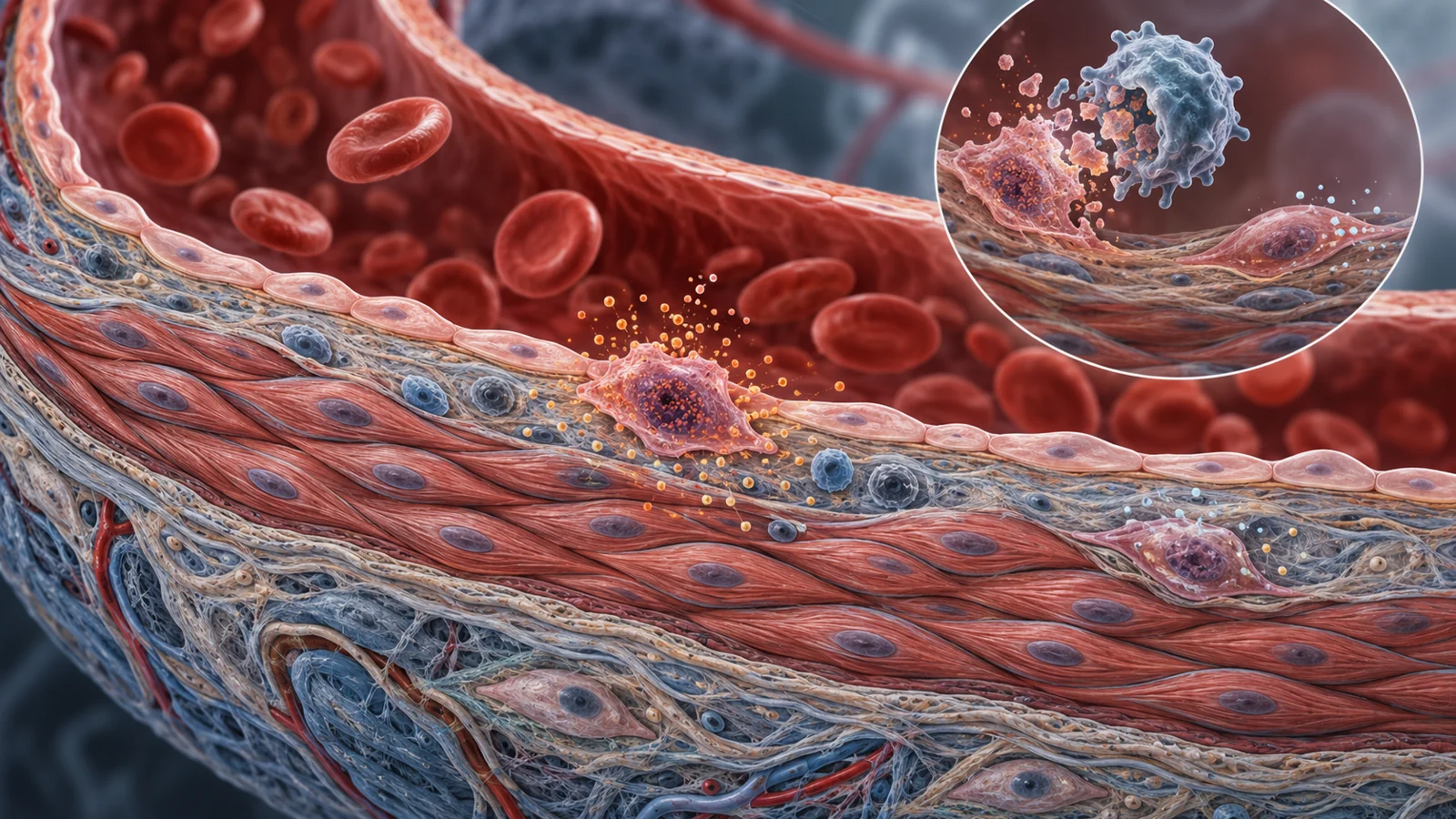
The inner lining of a blood vessel is made of endothelial cells. Healthy endothelial cells release nitric oxide, a molecule that helps the vessel relax. Nitric oxide also supports the barrier between blood and tissue, limits the display of adhesion molecules, and helps restrain coagulation. Senescent endothelial cells produce fewer protective signals and show more inflammatory and adhesion-related activity. They also lose some ability to divide and repair the vessel lining.
The review connects these changes with impaired blood-flow regulation. Reduced nitric oxide activity can limit vasodilation, while changes involving angiotensin II and endothelin-1 may contribute to higher blood pressure. Senescent endothelial cells also produce more reactive oxygen species and SASP factors. Because the endothelium acts partly like an endocrine tissue, its dysfunction may influence more than the small area where the cells reside. The review also discusses an association between reduced vasodilation from senescent endothelial cells and heart failure with preserved ejection fraction.
The muscular layer beneath the lining contains vascular smooth muscle cells, which help control vessel diameter and maintain the artery’s structure. As these cells become senescent, they can release inflammatory signals such as interleukins and tumor necrosis factor-alpha. Their contractile machinery becomes less effective, and their responses to signals from endothelial cells can change. Those alterations may impair blood-flow control and contribute to arterial stiffness.
Senescent vascular smooth muscle cells may also produce more matrix-degrading proteases and less collagen. In an atherosclerotic plaque, a weaker collagen structure can make the plaque more vulnerable to rupture. Oxidative stress and inflammation can additionally push these cells toward producing factors associated with vascular calcification. These are different routes by which a small population of altered cells could affect the mechanical and inflammatory behavior of a larger vessel wall.
Two ways of targeting the problem
Senolytics are drugs intended to eliminate susceptible senescent cells. They do this by interfering with survival systems that senescent cells use to resist apoptosis, or programmed cell death. The review describes these systems as senescent cell anti-apoptotic pathways. Different senescent cell types can depend on different survival signals, including pathways involving SRC kinases, PI3K, BCL-xL, mitochondrial processes, or p21-related regulation.
That variety matters. A drug that removes one vulnerable population may leave another population untouched, and a cell that looks similar by one laboratory marker may rely on a different survival pathway. Dasatinib and quercetin, often discussed together as D+Q, are examples of a senolytic combination examined in the research summarized by the review. Their effects are not evidence that the same combination is a universal cardiovascular treatment.
Senomorphics take a different approach. They do not directly remove senescent cells. Instead, they try to alter harmful parts of the SASP or other senescence-associated activity. Possible targets discussed in the review include NF-kappa B, Janus kinase and STAT signaling, mTOR, p38 MAPK, mitochondrial pathways, and NAD metabolism. Medications such as metformin, rapamycin, and ruxolitinib are discussed for senomorphic effects in particular contexts, but that discussion is not a recommendation to use them for cardiovascular disease.
The distinction is useful because reducing a damaging secretome and removing its source are not interchangeable actions. A senomorphic treatment might quiet inflammatory signaling while leaving the cell in place. A senolytic might reduce the source but also affect cells with different roles in different tissues. Either strategy would need to be evaluated by what happens to vessel function, cardiac structure, and whole-body safety—not simply by whether a marker goes down.
What the mouse studies actually measured
In aged mice, the review reports that D+Q improved vasorelaxation dependent on both endothelial cells and vascular smooth muscle cells. The finding connects the intervention with a functional measure: the ability of blood vessels to relax. It is more informative than showing only that a senescence-associated marker changed, because vessel relaxation is part of how the cardiovascular system regulates blood flow. Even so, an improved response in aged mice does not establish a human benefit or show that the treatment can prevent cardiovascular events.
A separate example involved navitoclax in a mouse model of ischemia–reperfusion injury. The review reports reduced myocardial hypertrophy and fibrosis, improved left ventricular ejection fraction, and increased myocardial vascularization. Here the experimental setting was a heart exposed to a period of interrupted blood flow followed by restoration, and the measured outcomes concerned adverse remodeling and pumping function. This is a different model, intervention, and set of endpoints from the aged-mouse vessel-relaxation experiment.
The review also describes laboratory work in which human induced-pluripotent-stem-cell-derived cardiomyocytes and endothelial cells were exposed to senescent cells. The co-culture setting was associated with lower cell survival and activity, while D+Q improved several cellular measures, including survival and endothelial migration and tube formation. Such experiments can help connect secreted factors with cell behavior, but they are not a substitute for treatment in people with cardiovascular disease.
Why less senescence is not always better
Senescence has a protective side. When a cell suffers DNA damage, permanent growth arrest can prevent uncontrolled proliferation. If the body could simply block cells from entering senescence, damaged cells might continue dividing. The review therefore distinguishes preventing senescence formation from eliminating selected senescent cells that already exist. Interfering broadly with pathways such as p16 or p53-p21 could weaken an important barrier against tumor formation, and the risks of that approach should not be transferred automatically to every senolytic strategy.
Fibrosis also needs a more precise interpretation. Excessive fibrosis can stiffen tissue and impair cardiac function, which makes it an attractive target in some disease settings. Yet after a myocardial infarction, scar tissue can replace dead muscle and help preserve the heart wall. The fibrous cap of an atherosclerotic plaque can likewise help prevent rupture. A treatment that reduces all fibrosis would not necessarily be beneficial.
The review notes that dysfunctional senescent fibroblasts may be a more selective target than the normal repair process carried out by non-senescent fibroblasts. That possibility is not a guarantee of long-term safety. It is a reason to distinguish pathological remodeling from repair that the cardiovascular system still needs.
From a marker in a sample to a treatment that works
Identifying senescent cells is itself difficult. The review lists markers including p16, p21, DNA-damage signals, p38 MAPK activity, telomere-associated foci, and senescence-associated beta-galactosidase. None is fully sensitive and specific. SASP factors measured in blood or tissue can provide additional information, but individual factors can also be released by other cell types. Composite panels may give a better estimate of senescent cell burden than any single measurement.
A lower marker level would therefore be an intermediate observation, not proof that cardiovascular health has improved. A convincing treatment assessment would need to connect cell targeting with meaningful function: better vascular relaxation, safer remodeling after injury, preserved tissue integrity, and outcomes that remain favorable over time. The review identifies the long-term effects of systemic senescent-cell elimination as an unresolved challenge. Senomorphic drugs also need evaluation for effects on local tissue balance and immune function.
Human senolytic research discussed in the review included work in idiopathic pulmonary fibrosis and other diseases or geriatric syndromes. Those studies may inform the broader field, but lung-disease findings do not establish cardiovascular efficacy. The supplied evidence also does not provide a validated test for selecting cardiovascular patients, a universally effective drug, or a safe treatment schedule.
What this could change
The possibility is a more selective form of cardiovascular treatment for older adults: one that helps arteries relax more effectively and reduces damaging remodeling after heart injury by targeting harmful senescent-cell activity. Reaching that point would require the animal findings to translate into meaningful human outcomes, while selective treatment preserves the growth-arrest, scar-forming, and repair functions that protect the heart and arteries.
That conditional path is the important part of the story. Senescent cells may offer an additional target beyond familiar cardiovascular risk factors, but the therapeutic value will depend on which cells are targeted, which signals are changed, and whether the surrounding tissue remains able to repair itself safely.
Key Sources
- Primary source: Senescent Cells: A Therapeutic Target in Cardiovascular Diseases.
DOI or Journal Page · Review or Meta-Analysis · 2023-05-02
Keep exploring