
Research Signal – Source date 2021-09-21
A cell that stops dividing sounds like a useful barrier against cancer. Uncontrolled division is one of cancer’s defining problems, so forcing damaged or malignant cells into a lasting growth arrest might seem like a way to contain disease. But growth arrest does not mean that the cell has become inert.
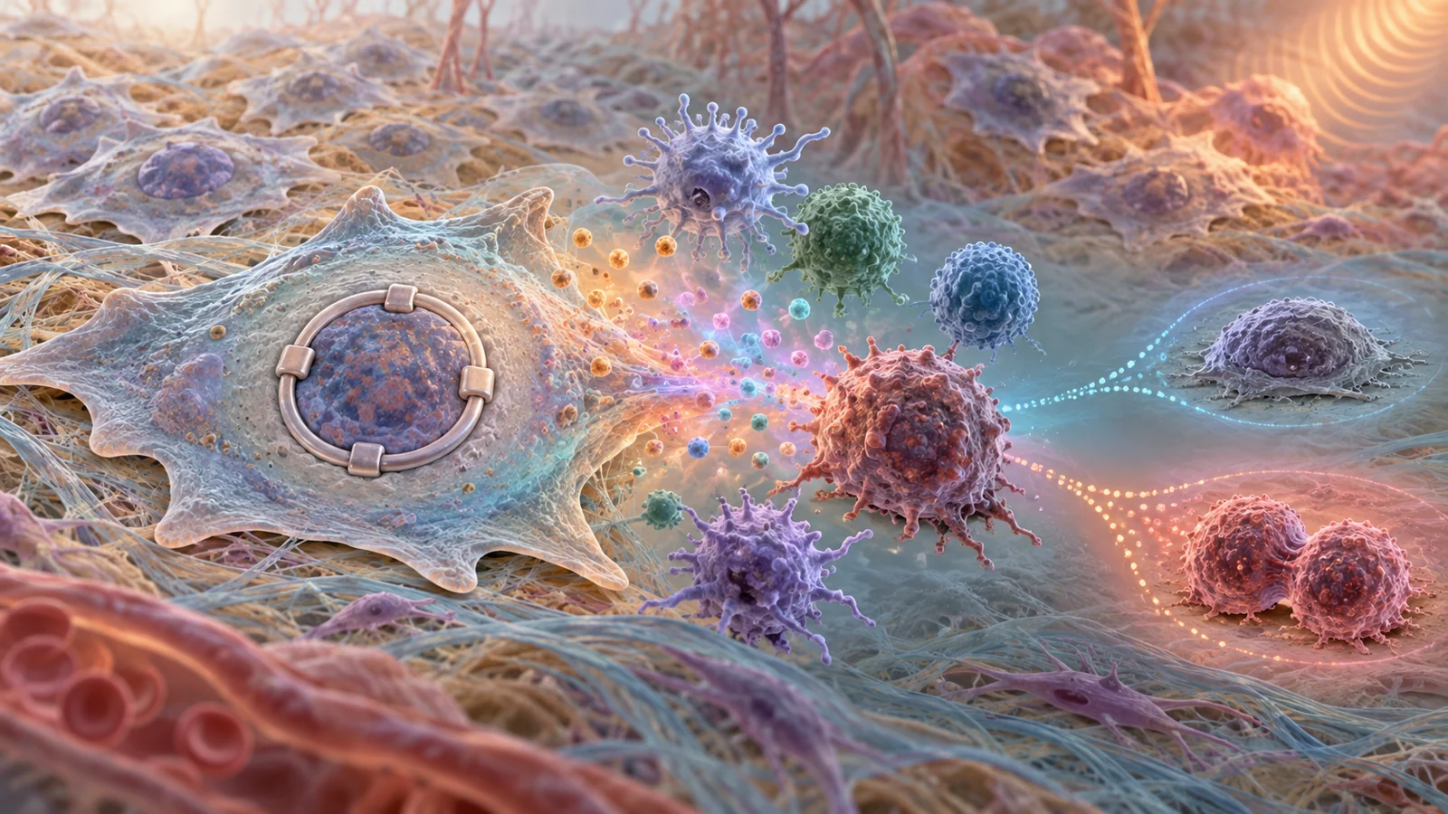
Senescent cells remain metabolically active. They can release inflammatory agents, growth factors and proteases that alter nearby cells and the tissue around them. This collection of secreted substances is known as the senescence-associated secretory phenotype, or SASP. Its contents vary with the cell’s tissue of origin, the stress that produced senescence and the time since that stress occurred.
A review published on September 21, 2021, brought together evidence from cell experiments, animal models and human observations to examine how this biology might connect aging, cancer treatment and tumor progression. The review’s central concern is not that senescence always helps or always harms cancer. It is that a growth-arrested cell may still influence what happens around it, while some cancer cells in experimental systems may later escape the arrested state.
A growth arrest that is not biological silence
Cellular senescence is a stress response that generally involves sustained exit from the cell cycle. Telomere shortening, DNA damage, oncogene activity, chemotherapy and irradiation can all trigger pathways associated with senescence. These stresses converge on mechanisms that stop the cell from entering another round of division.
The arrested cell, however, continues to function in other ways. It can change its metabolism and release a mixture of signals. Some of those signals reinforce senescence in neighboring cells, but others can encourage inflammation, alter tissue structure or change the behavior of cancer cells. The result depends on which cells are present, what caused senescence and how the local environment responds.
This matters because cancer progression is not controlled only by the division rate of the cancer cells themselves. Surrounding fibroblasts, immune cells, blood vessels and structural tissue can influence whether malignant cells remain contained, invade nearby tissue or withstand treatment. A senescent cell can therefore affect cancer without directly becoming a dividing cancer cell.
Aging and treatment create related but different senescent populations
During ordinary aging, senescent cells gradually accumulate. The review describes this as a balance between their production and their clearance: as the number produced rises or the ability to remove them falls, the burden increases. Telomere erosion, DNA damage, epigenetic disruption and mitochondrial dysfunction are among the processes associated with age-related senescence. Senescence markers have also been observed in older people, alongside higher levels of inflammatory factors such as interleukin-6.
Chronological age is not the only route to this state. Werner syndrome, a rare inherited premature-aging disorder, provides an example of how accelerated aging biology can appear earlier in life. Cells from affected patients can undergo premature replicative senescence, and senescent cells produced by reducing WRN activity in laboratory experiments released signals that increased proliferation and other tumor-promoting properties in recipient cells. This is evidence from a disorder-specific and experimental setting, not a measure of cancer risk for people with ordinary aging.
Chemotherapy creates another context. Treatment can induce senescence in cancer cells and in noncancerous cells that experience DNA-damaging stress, a process often called therapy-induced senescence. Senescence markers have been reported in patients treated for breast cancer long after chemotherapy ended, but such observations do not show that secreted factors caused an individual patient’s cancer to progress.
The therapeutic context changes the question. A senescent cell created during aging may have been present as part of a long accumulation. A senescent cell created by treatment may remain after the drug has stopped, carrying a different history and potentially a different secretory profile. The review emphasizes that these profiles are heterogeneous rather than a single, universal recipe.
The messages can act on cancer cells or their surroundings
The review describes two broad destinations for senescence-associated secretions. They can act directly on cancer cells, or they can reshape the tumor microenvironment that supports and restrains those cells.
One way to make the first route tangible is to look at conditioned medium. In cell experiments, fluid collected from senescent fibroblasts stimulated excessive proliferation and progression in premalignant and malignant epithelial cells. Secretions from aged fibroblasts also promoted therapeutic resistance in melanoma cells by supporting local invasion and metastasis-related behavior. Similar experiments with senescent mesenchymal cells increased the proliferation of breast cancer cells.
The second route runs through the surrounding tissue. In a mouse prostate cancer model, a senescence-associated environment was linked with immunosuppressive features, including infiltration by myeloid-derived suppressor cells and an absence of natural killer cells in the model described by the review. Other experiments connected interleukin-6 from senescent skin stroma with the recruitment and expansion of immunosuppressive myeloid cells. Interleukin-8 from senescent cancer-associated fibroblasts promoted invasion and metastasis in mouse models of pancreatic cancer.
These findings describe biological behavior in cells and animals. They do not quantify how much human cancer incidence or relapse is attributable to senescence-associated inflammation. They do show why the effect of a senescent cell cannot be judged only by asking whether that cell is dividing. Its released signals may change the conditions in which other cells survive, move or respond to treatment.
Relapse involves more than one possible route
Secretions from senescent cells are one possible route to a more aggressive tumor environment. Another is the behavior of the arrested cancer cells themselves. The review discusses experimental systems in which cancer cells induced to senesce after chemotherapy later resumed proliferation or formed tumors when implanted in mice.
Several examples involve lung, colon, breast, liver and lymphoma models. In one set of experiments, cells released from doxorubicin-induced senescence showed more aggressive growth potential than cells exposed to chemotherapy without entering senescence. In liver cancer cells, treatment-associated senescence coincided with higher expression of reprogramming and stemness-related genes and greater tumor-forming ability in vivo. These observations suggest that an arrested state can sometimes be temporary or reversible in cancer cells under particular experimental conditions.
The review also separates a temporary senescent-like state from classic senescence. In an acute myeloid leukemia model, chemotherapy produced cells with a temporary growth arrest and an inflammatory program that may have helped them withstand treatment, providing one possible cellular basis for relapse after therapy ended. A temporary pause in division is not equivalent to a permanently arrested cell, even if both states release similar signals.
The two routes can overlap, but they are not interchangeable. A cancer cell might resume division after senescence, while a neighboring fibroblast remains arrested and changes the environment through its secretions. Conversely, a cancer cell could remain growth-arrested while secreted factors alter other cells. In some lymphoma and liver cancer examples, the review explicitly notes that the specific contribution of secretion to the aggressive behavior was not investigated. The experiments therefore support a distinction between escape from arrest and effects transmitted through the SASP, without proving that one mechanism explains every relapse-like outcome.
Three ways researchers might separate harm from protection
The therapeutic ideas discussed in the review operate at different biological levels. The broadest is senolysis: selectively removing senescent cells. In mouse studies, clearing senescent cells reduced age-associated phenotypes. In another mouse model, eliminating doxorubicin-treated senescent fibroblasts prevented or delayed relapse and metastasis and reduced treatment-associated cardiac dysfunction and bone-marrow suppression. Navitoclax also increased the effectiveness of some chemotherapy regimens in models of lymphoma and multiple myeloma. These are experimental findings, not evidence that senolytic treatment prevents relapse or improves survival in cancer patients.
A second approach is to suppress the machinery that produces the secretory phenotype while leaving growth arrest in place. The review discusses the interleukin-1 pathway as an upstream regulator of inflammatory SASP production. It also describes how mTOR inhibition or rapamycin reduced parts of the SASP in several cellular senescence settings without reversing cell-cycle exit. This approach could, in principle, preserve the useful barrier to proliferation while limiting inflammatory activity, but the effects remain dependent on the tissue, stimulus and specific pathway involved.
A third approach is narrower: block selected secreted factors that appear to support cancer progression. The review cites experimental examples involving CCL2 and amphiregulin, where targeting those factors reduced colorectal cancer progression or treatment resistance in the systems described. A targeted intervention might avoid removing every senescent cell or suppressing every component of its secretory program. It might also miss harmful factors that have not been identified, or interfere with signals that serve a protective function in another setting.
Choosing among these strategies is difficult because senescence is not one uniform state. The secretory profile can change across tissues and over time, and the same broad response may restrain one process while supporting another. Removing many senescent cells at once could also create tissue problems, a concern raised in the review when discussing the possible consequences of extensive clearance in aged or diseased organs. The practical goal would therefore be more precise than simply eliminating senescence: preserve growth arrest where it helps, limit harmful signaling where it matters, and establish that the balance is safe and beneficial in people.
What this could mean for lasting cancer control
The prospect raised by this 2021 review is a more durable form of cancer treatment that accounts for what remains after therapy has stopped. Future treatments might combine the useful effect of keeping damaged or malignant cells from dividing with a way to reduce inflammatory signals from senescent cells or prevent surviving cancer cells from escaping arrest. That could make a treatment response more durable by addressing both the cells left behind and the environment they help create.
Reaching that possibility would require context-specific targeting and demonstrated benefit in people. The review does not establish a universal secretory profile, a single intervention or a patient-level method for predicting relapse from senescence markers. Its contribution is to connect aging-associated accumulation and treatment-induced senescence, showing why lasting cancer control may depend on separating the protective and harmful functions of a state that looks quiet from the outside but remains biologically active.
Key Sources
- Primary source: The Contribution of Physiological and Accelerated Aging to Cancer Progression Through Senescence-Induced Inflammation.
DOI or Journal Page · Review or Meta-Analysis · 2021-09-21
Keep exploring