
Research Signal – Source date 2021-10-18
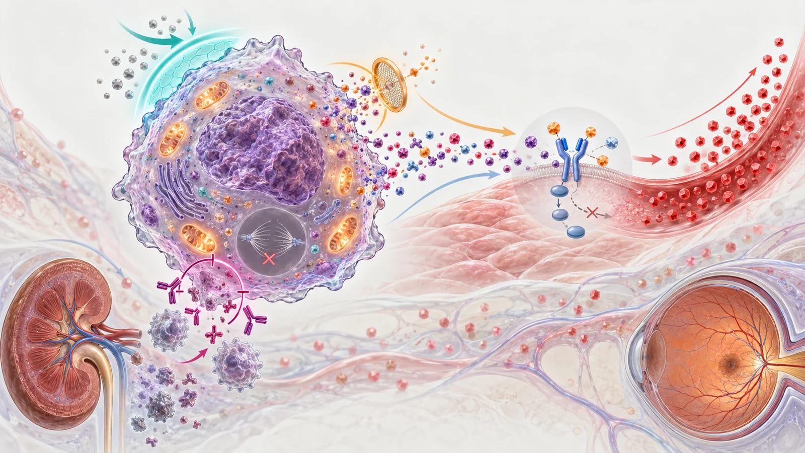
Cells that have entered senescence stop dividing, but they do not necessarily become biologically silent. Instead, many remain active and release a changing mixture of molecules known as the senescence-associated secretory phenotype, or SASP. These signals can influence nearby cells, immune responses and tissue function.
That combination creates an important tension. Senescence can protect young organisms by stopping damaged or potentially cancerous cells from multiplying. It also appears temporarily during development and tissue repair, where secreted factors can help shape structures or support healing. But when senescent cells persist or accumulate in the wrong setting, their signals may contribute to inflammation and metabolic dysfunction.
A review published on October 18, 2021, examined how metabolism and senescence affect one another. Its central idea is not that every senescent cell behaves the same way, or that one metabolic pathway explains aging. It is that cellular energy balance, nutrient handling and stress responses can influence both the formation of senescent cells and what those cells do afterward.
When a stopped cell keeps sending signals
Cellular senescence is usually described as a permanent halt to cell division accompanied by broader changes in cell behavior. The arrest may involve regulators such as p16INK4a and p21CIP1, but neither regulator appears in every senescent cell. The same variability applies to commonly used markers, including senescence-associated beta-galactosidase, reduced lamin B1 and the movement or secretion of HMGB1.
This matters because a marker is not the same thing as a complete diagnosis. A tissue may show a change in one indicator without establishing that all relevant cells are senescent, that the cells have been eliminated, or that the tissue has become healthier. The review therefore emphasizes using multiple markers when studying senescence in living tissues.
The biological effects are also context-dependent. DNA damage, activated oncogenes and mitochondrial dysfunction can induce senescence in cultured cells and animal models, but identifying what initiates senescence during natural aging is more difficult. Senescent cells are often uncommon even in old or diseased tissues, and they can reprogramme their metabolism to remain alive while maintaining a complex secretory state.
A metabolic loop can amplify diabetic stress
The review brings several diabetes-related findings into one disease model. Senescent cells outside the pancreas can contribute to insulin resistance in obese mice. Insulin resistance can then increase pressure on pancreatic beta cells, while metabolic stress and high blood glucose can promote senescence in vulnerable cells. Senescence associated with elevated glucose has also been linked in the review to kidney and retinal complications in experimental diabetes.
The sequence is best understood as a reinforcing process rather than a single linear cause. Peripheral senescence may impair metabolic control; worsening metabolic control may encourage further senescence; and affected organs may then accumulate additional dysfunction. This is an integrated interpretation of findings across tissues and experiments, not evidence that senescence alone explains diabetes.
One mouse finding shows why the biology is more complicated than a simple harmful-cell story. When p16INK4a was increased specifically in pancreatic beta cells, insulin secretion rose. In that setting, a senescence-associated state may have represented a local compensation for high blood glucose, even though senescent cells elsewhere could contribute to insulin resistance and disease progression. The same broad cellular program can therefore have different consequences depending on its location and stage.
The practical implication is that an intervention aimed at senescence must specify what it is trying to change. Preventing new senescent cells, reducing the secretions of existing cells and removing those cells are related strategies, but they are not interchangeable biological operations.
NAD+ changes can push different parts of the phenotype
NAD+ is involved in energy metabolism and also serves as a cofactor for enzymes such as PARP and sirtuins. Its relationship with senescence illustrates why a metabolic intervention cannot be judged from one readout alone.
Mitochondrial dysfunction can cause a senescence phenotype in which the balance between NADH and NAD+ shifts because mitochondria are less able to oxidize NADH. The resulting change in the NAD+/NADH ratio can inhibit GAPDH, reduce ATP availability, activate AMPK and contribute to cell-cycle arrest. This form of mitochondrial dysfunction-associated senescence has a distinct secretory profile that lacks some of the strongly pro-inflammatory features seen in other contexts.
Total NAD+ abundance introduces a different layer. Lower NAD+ can promote senescence by weakening processes that protect against cellular stress, and NAD+ levels decline with age in many tissues. Yet once senescence is established, the review describes senescent cells with increased NAMPT and NAD+. That state can reduce AMPK and p53 activity while increasing p38MAPK and NF-kB activity, supporting the release of inflammatory SASP factors.
The result is a state-dependent relationship. A change that might influence whether a cell enters growth arrest does not automatically have the same effect on the secretions of a cell that is already senescent. These findings concern cellular mechanisms and do not establish the clinical benefits or risks of NAD+ supplements.
A damaged lysosome can become a liability
The review also describes a more selective route from altered metabolism to potential cell removal. Many senescent cells have dysfunctional lysosomes, the compartments that help digest and recycle cellular material. Increased lysosomal permeability can reduce acidity inside the lysosome while contributing to acidification of the surrounding cell interior.
Senescent cells appear to compensate by increasing glutaminolysis, the breakdown of glutamine. Higher activity of glutaminase 1 produces ammonia, which helps neutralize excess acidity in the cytosol. In experimental systems, blocking glutaminase 1 killed senescent cells in culture and in animals and improved several indicators of age-related organ dysfunction.
This is not the same as broadly correcting metabolism across the body. It is an example of a survival dependency: a defect that helps create cellular stress may also force senescent cells to rely on a compensatory pathway. Inhibiting that pathway can become a research strategy for selective elimination, although the review does not establish a validated human treatment or dosing plan.
Prevention, suppression and removal pursue different goals
The first strategy is to slow the formation of senescent cells. In diabetic mice, lowering glucose with insulin or reducing glucose transport into kidney proximal tubule cells with an SGLT2 inhibitor prevented an increase in senescence there. Acarbose, which reduces intestinal carbohydrate absorption, also prevented diet-associated senescence linked to atherosclerosis in rabbits. These findings support glucose control as an experimental prevention route, not as proof that a particular human regimen prevents senescence-related complications.
A second strategy leaves senescent cells in place but reduces their harmful activity. Metformin is the review’s best-characterized metabolic example. In cell and animal models, it opposed several senescence responses, lowered parts of the pro-inflammatory SASP and interfered with NF-kB activation. Exercise likewise prevented some senescence and SASP changes in mouse adipose tissue and liver exposed to a poor diet. In humans, physical activity was associated with lower markers of senescence in endothelial and immune cells, but those associations do not by themselves establish that exercise caused the change or that the change improved a clinical outcome.
The third strategy is senolysis, the removal of senescent cells. The review describes experimental senolytic approaches that improved blood-glucose measures or diabetes-related complications in animal studies. Removing cells differs from preventing their formation or quieting their secretions. The authors note that removal could potentially be intermittent, while preventive and suppressive approaches would require continued exposure or adherence to maintain their effects. That proposed scheduling advantage is a practical hypothesis, not a validated human dosing schedule.
These strategies may eventually be combined, but the review does not identify a clinically superior option. Their effects could also differ across organs, cell types and stages of disease. A treatment that reduces a marker or a SASP factor may be addressing a different problem from one that eliminates a cell.
Measurement changes the meaning of a result
Senescence research is especially vulnerable to overinterpretation because the state has no single universal marker. A convincing assessment usually needs several independent indicators of growth arrest, cell identity and secretory or structural change. Even then, the biological meaning of a measurement depends on the tissue and the question being asked.
A lower level of senescence-associated beta-galactosidase, for example, could reflect altered marker expression rather than the disappearance of senescent cells. A reduction in inflammatory signals could mean that cells are secreting less, not that they have been removed. Neither result alone demonstrates reversal of organismal aging or improved patient health.
The distinction between experimental induction and natural aging is equally important. Much of the mechanistic evidence comes from cultured cells or animal models exposed to defined stresses. Single-cell profiling was emerging as a way to identify which drivers matter during natural aging and age-related disease, but the review did not turn those unresolved questions into a clinical answer.
Human observations can still help locate the relevant biology. Associations between physical activity and lower senescence markers in human endothelial or leukocyte populations suggest that the relationship between lifestyle, metabolism and cellular state may matter beyond laboratory models. They do not establish the direction of causation, the responsible pathway or a treatment effect. Careful interpretation must keep those questions separate.
What this could mean for diabetes complications
The most concrete prospective benefit would be protecting people with metabolic disease from the progressive kidney and retinal damage associated with diabetes. The review’s model suggests several possible routes: reduce the metabolic conditions that help create senescent cells, suppress the harmful signals released by cells that remain, or remove vulnerable senescent populations after they form.
That possibility depends on translating the reviewed mechanisms into interventions that are safe, effective in humans and selective enough to preserve useful senescence responses involved in cancer protection and repair. The findings do not yet choose a winning strategy. They show why diabetic complications could become a testing ground for treatments that address cellular state and metabolic feedback together, provided the experimental distinction between prevention, suppression and removal survives clinical evaluation.
Key Sources
- Primary source: The metabolic roots of senescence: mechanisms and opportunities for intervention.
DOI or Journal Page · Review or Meta-Analysis · 2021-10-18
Keep exploring