
Research Signal – Source date 2026-03-16
In a mouse study published March 16, 2026, animals given resveratrol performed better on a memory-related test after an accelerated-aging regimen. Their brains also showed lower levels of several markers associated with cell senescence and inflammation. Separate experiments in cultured microglial cells showed reduced activity in the cGAS-STING pathway, an intracellular DNA alarm system, after resveratrol treatment.
The findings connect a behavioral result with changes in brain tissue and cell signaling. They do not show that resveratrol clears senescent brain cells, or that it protects memory in people. The model, measurements, and cell experiments help explain what the researchers observed—and where the interpretation must stop.
The memory result came from an induced-aging mouse model
The researchers assigned 36 male mice to three groups: a control group, a D-galactose group, and a group given resveratrol during the final three weeks of D-galactose dosing. D-galactose was injected daily for eight weeks to induce features of accelerated aging. During the final three weeks, the treatment group received resveratrol by mouth each day; the other groups received the corresponding vehicle treatment.
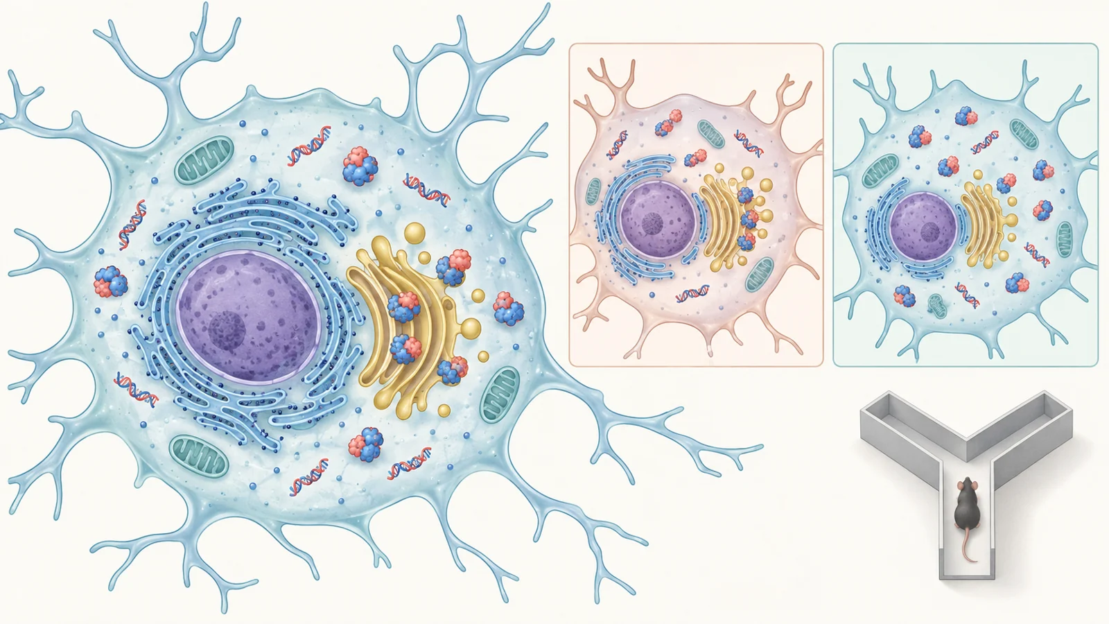
After the regimen, the team used behavioral tests to assess movement, anxiety-related behavior, and memory-related performance. In a Y-shaped maze, mice first explored two arms, then later had access to those familiar arms and a third, new one. The researchers compared entries into the new arm with entries into the familiar arms. This test uses a mouse’s tendency to explore a novel space as a measure related to recognition memory; it is not a direct account of a person remembering an event.
The D-galactose group showed impaired cognitive performance compared with controls, while resveratrol-treated mice performed better on the memory-related assessment. An open-field test and a rotarod test provided additional behavioral context, including spontaneous movement and motor coordination. Those measures matter because activity or motor differences can affect how an animal performs in a maze. The reported behavioral comparison supports an improvement in this mouse model, not a general claim about memory across settings or species.
Brain staining and inflammatory transcripts changed too
The study also examined brain tissue, including the hippocampus and cerebral cortex. D-galactose-treated mice had more senescence-associated beta-galactosidase staining in these regions than controls. Resveratrol-treated mice had less. The researchers also found lower staining signals associated with oxidative DNA damage and lower measures of senescence markers in microglia, the brain’s resident immune cells.
These are measurements of features associated with senescence. In particular, the team used staining and marker co-localization to assess whether microglia displayed senescence-related signals. A reduction in those signals is evidence that the measured phenotype changed; it does not show that resveratrol selectively removed senescent microglia. Establishing cell clearance would require evidence about the fate and number of those cells, not just lower marker levels.
The researchers measured inflammatory transcripts in the cortex and hippocampus as well. D-galactose exposure raised several factors associated with the senescence-associated secretory phenotype, or SASP, including Cxcl-10, Ccl-2, Ifi-44, Il-1β, and Il-18. Resveratrol treatment lowered their measured expression. These RNA measurements indicate changes in gene expression in the sampled tissue; they are not themselves a direct measurement of every inflammatory protein or of the brain’s overall immune state.
How a DNA alarm can sustain inflammation
The proposed pathway begins when DNA appears in the cell’s cytoplasm, outside the nucleus where most of the cell’s genetic material is normally kept. Damaged nuclear or mitochondrial DNA can provide this misplaced material. The sensor cGAS can detect cytoplasmic DNA and produce a messenger molecule that activates STING. STING then helps relay an immune response, including signals that involve TBK1 and inflammatory gene expression.
A short-lived response to foreign or misplaced DNA can be useful for defense. The concern in aging is that persistent activation may keep inflammatory signaling switched on. In microglia, that could contribute to a senescence-associated state and continued release of inflammatory factors. This is the biological rationale for examining cGAS-STING in the study. It is broader context for interpreting the experiments, not a direct measurement of every step in naturally aging human microglia.
Several assays pointed to lower pathway activity
In brain samples from the mice, the researchers measured phosphorylated STING and phosphorylated TBK1, proteins associated with activated pathway signaling. Both were elevated in the D-galactose group and lower after resveratrol treatment in the cortex and hippocampus. These protein measurements add a pathway-related readout alongside the tissue staining and inflammatory transcripts.
The team then used cultured microglial cell lines to examine the pathway more directly. In BV-2 mouse microglial cells, they used double-stranded DNA to stimulate cellular DNA-sensing responses and induce senescence-associated features. Resveratrol reduced measured senescence markers and inflammatory factors in these stimulated cells. It also reduced phosphorylation of STING and TBK1. The methods also describe culturing HMC-3 human microglial cells, but these cell lines remain laboratory models rather than intact human brain tissue.
RNA sequencing in the BV-2 cells provided a broader view of gene activity. DNA stimulation increased expression patterns associated with cytosolic DNA sensing and inflammation; after resveratrol treatment, those pathway signatures were less prominent. Sequencing measures patterns across many genes, while protein assays measure particular signaling components and brain-tissue tests show changes in the animal. Their agreement makes the pathway a plausible part of the response. It does not prove that cGAS-STING inhibition caused the memory improvement: the study did not isolate that pathway as the sole route from resveratrol to behavior.
STING’s route to the Golgi was a distinctive finding
STING is normally associated with membranes in the endoplasmic reticulum, an internal cell compartment. After activation, it moves toward the Golgi apparatus, where it can organize downstream signaling. The researchers tracked STING’s location in cultured BV-2 cells by comparing its signal with GM130, a marker of the Golgi.
After double-stranded DNA stimulation, more STING signal overlapped with the Golgi marker. Resveratrol pretreatment reduced that overlap, leaving STING more diffusely distributed in the cells. The researchers also tested H-151, a STING inhibitor. Under the reported conditions, H-151 did not produce the same change in STING localization. Resveratrol’s effect on this movement therefore differed from the comparator’s effect in this assay.
The team further stimulated cells with cGAMP, which activates STING while bypassing the DNA-sensing step upstream. Resveratrol still reduced STING’s movement toward the Golgi. This narrows the possible location of its influence in the cell experiments, but it does not identify a direct molecular target. The results do not establish that resveratrol binds STING or that altered trafficking alone explains the memory result. Effects on cell membranes, oxidative stress, or other regulators remain possible explanations raised by the study’s authors, not resolved mechanisms.
The model leaves natural aging and human benefit open
D-galactose exposure is an experimental way to produce aging-associated changes in mice. It does not reproduce the full course of natural aging, with its varied biological changes over time. The behavioral result therefore needs confirmation in naturally aged animals before it can be assumed to apply more broadly to aging brains.
The cell experiments answer a different question from the animal tests. Cultured microglial lines let researchers examine signaling and STING location under controlled stimulation, but they do not reproduce the interactions among brain cells and tissues in an intact animal. Meanwhile, the mouse study tested one treatment schedule in male animals. It cannot establish a useful dose, safety, or memory benefit for people.
The most defensible reading is that resveratrol was associated with improved memory-related performance and lower senescence-associated and inflammatory measurements in this induced-aging mouse model. The cell experiments offer a possible pathway explanation, especially the change in STING trafficking, while leaving the exact mechanism and its contribution to behavior unresolved.
What quieter brain immune cells could mean for memory
If the pathway findings hold up in natural aging and the behavioral effect proves reproducible, a treatment that limits persistent DNA-triggered inflammation could eventually help preserve memory as the brain ages. That possibility depends on establishing how resveratrol acts, whether the effect can be achieved safely in people, and whether changes in microglial signaling translate into meaningful cognitive benefit. For now, the study offers a mechanistic lead in mice and cells, not evidence that a plant-derived compound protects human memory.
Key Sources
- Primary source: Resveratrol Mitigates Age-Associated Cognitive Decline via Inhibition of cGAS-STING-Mediated Microglial Senescence.
DOI or Journal Page · Peer-Reviewed Paper · 2026-03-16
Keep exploring