
Research Signal – Source date 2021-10-04
Keeping a transplanted kidney working for years is a central challenge after transplantation. The kidney may be functioning, yet the tissue remains exposed to surgical stress, immune activity, infections, rejection episodes and the effects of immunosuppressive treatment.
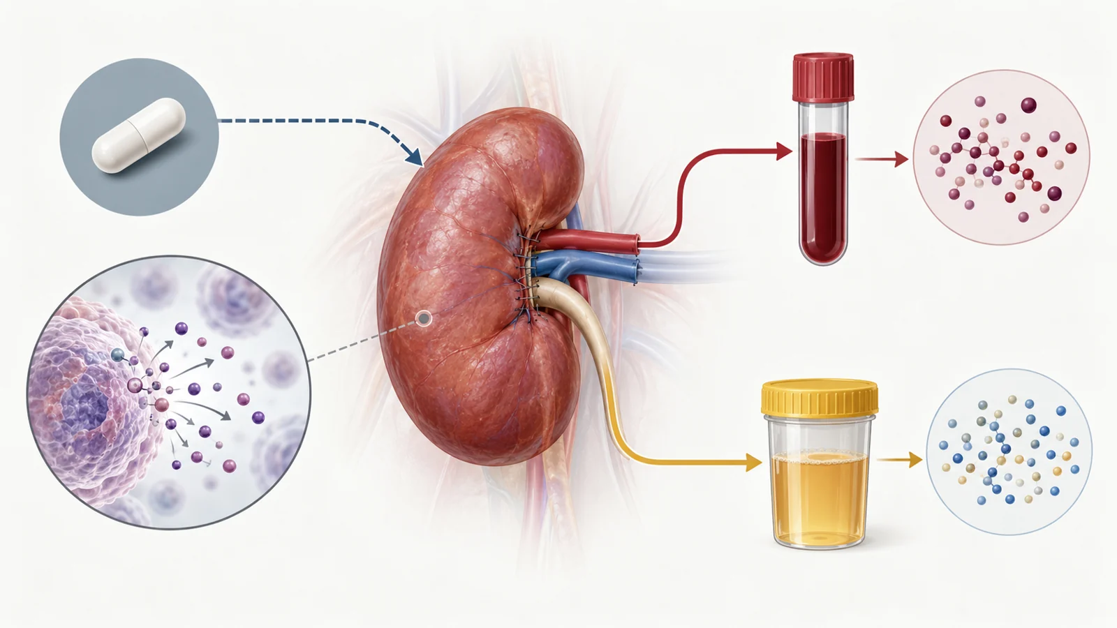
One possible contributor is cellular senescence. A senescent cell has permanently stopped dividing, but it may remain biologically active. It can release inflammatory cytokines, growth factors, chemokines and enzymes into its surroundings. Researchers call this collection of secretions the senescence-associated secretory phenotype, or SASP. These substances can influence neighboring cells and promote further tissue stress.
The October 4, 2021 ORENTRA paper asked whether marine omega-3 fatty acids could alter some of those circulating secretions in people who had recently received a kidney transplant. It was an exploratory substudy of a randomized human trial. The question was whether omega-3s changed a selected set of proteins associated with senescence; those measurements could not establish reversal of cellular aging.
Inside the 44-week comparison
The study included 132 adult kidney transplant recipients in Norway. Each had a functioning graft eight weeks after transplantation, defined in the trial as an estimated glomerular filtration rate above 30 mL/min/1.73 m². Sixty-six participants were assigned to marine n-3 polyunsaturated fatty acids and 66 to an olive-oil placebo.
The trial was randomized, double-blind and placebo-controlled. Participants took three capsules daily for 44 weeks. The marine n-3 group received approximately 2.6 grams per day of the omega-3 fatty acids EPA and DHA; the comparison group received three olive-oil capsules. Both groups continued the local standard immunosuppressive regimen, including prednisolone, tacrolimus and mycophenolate.
The parent ORENTRA trial was organized around a kidney-function outcome: change in measured glomerular filtration rate, assessed using iohexol clearance. This substudy used the same trial setting for a different purpose. It examined a predefined panel of SASP-related proteins in plasma and urine. The sample size had been determined for the parent trial’s kidney-function endpoint, not specifically for this biomarker analysis.
Five blood findings stood out
After the intervention, the omega-3 group had more favorable between-group changes in five plasma measurements: granulocyte colony-stimulating factor, or G-CSF; interleukin 1 alpha, or IL-1α; macrophage inflammatory protein 1 alpha, or MIP-1α; matrix metalloproteinase 1, or MMP-1; and MMP-13.
The first three can be understood as inflammatory signaling proteins. MMP-1 and MMP-13 are enzymes involved in remodeling the material surrounding cells and tissues. The comparison concerned how measurements changed between the two groups. It should not be read as evidence that all five proteins fell from baseline in every person who took omega-3s.
MMP-13 provides a concrete example. Its median change was approximately zero in the marine n-3 group, while the control group showed a median increase. The between-group comparison was statistically significant. Similar group differences were reported for the other four highlighted plasma components.
The study measured 19 components overall. TGF-β1 was measured only in urine because its plasma measurement is especially vulnerable to confounding during blood collection and preparation. Among the urine measurements that could be analyzed, the investigators found no significant between-group differences. The blood and urine results therefore did not form a uniform pattern across every sample type or every protein.
The assay details change how the result reads
Several of the plasma measurements were difficult to quantify. Between 40% and 64% of plasma samples had concentrations below the assay’s detection limit for G-CSF, granulocyte-macrophage colony-stimulating factor, growth-related oncogene alpha, IL-1α and IL-1β. Many measurable values for these proteins were also close to the lower end of the assay’s standard curve. Two of the five proteins highlighted by the study, G-CSF and IL-1α, were among those affected by this issue.
That does not erase the reported group comparison, but it makes the numerical pattern less straightforward than a clean measurement of a plentiful substance. Results near a detection boundary can be more sensitive to how values are handled and to how much information the assay can distinguish at the low end.
The urine analysis had a different problem. A relatively large number of end-of-study urine samples were missing. Some were excluded because they showed signs of an ongoing urinary tract infection, while others were lost because of laboratory errors. Several urine components could not be measured reliably and were left out of the final analysis. The remaining nonsignificant urine comparison therefore cannot be treated as a complete survey of every possible urinary effect in the transplanted kidney.
There is also a biological limitation. The proteins in the panel are associated with processes that can occur around senescence, but they are not specific proof that senescent cells are present, increasing or being removed. The composition of the SASP varies by cell type, stage and trigger. Measuring a protein in plasma is therefore different from identifying senescent cells in a kidney biopsy.
MMP results require another layer of care. The measured concentration of MMP-1 or MMP-13 does not directly reveal how much enzyme is active or how much tissue remodeling is taking place. Tissue inhibitors can bind both active and inactive forms of MMPs, so a concentration change is not the same thing as a demonstrated change in enzyme activity.
The kidney outcome remained the crucial test
The parent trial found no significant difference between the marine n-3 and control groups in change in measured kidney function over 44 weeks. The substudy also found no association between changes in the five significant plasma components and changes in measured glomerular filtration rate or the percentage of fibrosis in kidney-graft biopsies.
This is the point at which the biological signal and the patient outcome separate. The blood findings suggest that supplementation influenced selected circulating proteins, but they did not show that the intervention preserved filtration or made the graft function better. Nor did they establish that the protein changes caused longer graft survival.
The parent trial did report a separate per-protocol finding: fibrosis scores in the control group increased while they remained stable in the marine n-3 group. That observation is relevant context, but it was not a demonstrated connection between the five highlighted proteins and tissue protection. The exploratory substudy did not supply that causal bridge.
Early recovery is not the same as long-term transplant care
The participants entered the trial eight weeks after transplantation, when recovery and adjustment to immunosuppression were still underway, and were followed until one year after transplantation. Bacterial or viral infections, acute rejection episodes and incidental malignancies occurred during follow-up and could influence circulating proteins. Their presence does not explain the treatment difference by itself, but it shows how many biological events can overlap with a biomarker measurement in this setting.
The participants also started with relatively high plasma phospholipid marine n-3 levels, above 6% on average in both groups. Levels rose in the supplementation group, providing evidence that the intervention increased exposure. Effects could differ in people beginning with lower omega-3 levels, so the result cannot automatically be transferred to every transplant population or to healthy adults.
Adverse events occurred in both trial groups. Gastrointestinal adverse events were the most frequent type of adverse event in both groups and the most common reason for withdrawal. The study was not designed to establish a general supplementation regimen for readers, and its 44-week experience does not provide a complete picture of long-term safety.
What this could change
A treatment that helps transplant recipients preserve kidney function for longer would offer a meaningful way to protect a graft while patients continue living with the demands of immunosuppression. Omega-3 supplementation could become part of that kind of adjunct approach only if the blood-protein pattern proves reproducible and is connected to evidence of senescence in graft tissue, followed by meaningful kidney outcomes in people further into long-term transplant care.
The ORENTRA substudy supplies an early biological lead rather than that clinical capability. Its value is in pointing toward a possible route: reduce harmful cellular signaling, confirm that the change reflects what is happening in the graft, and show that patients retain kidney function for longer. Until those links are demonstrated together, the result remains an intriguing measurement finding with a practical benefit still to be established.
Key Sources
- Primary source: Marine n-3 Polyunsaturated Fatty Acids and Cellular Senescence Markers in Incident Kidney Transplant Recipients: The Omega-3 Fatty Acids in Renal Transplantation (ORENTRA) Randomized Clinical Trial.
DOI or Journal Page · Peer-Reviewed Paper · 2021-10-04
Keep exploring