
Research Signal – Source date 2025-10-14
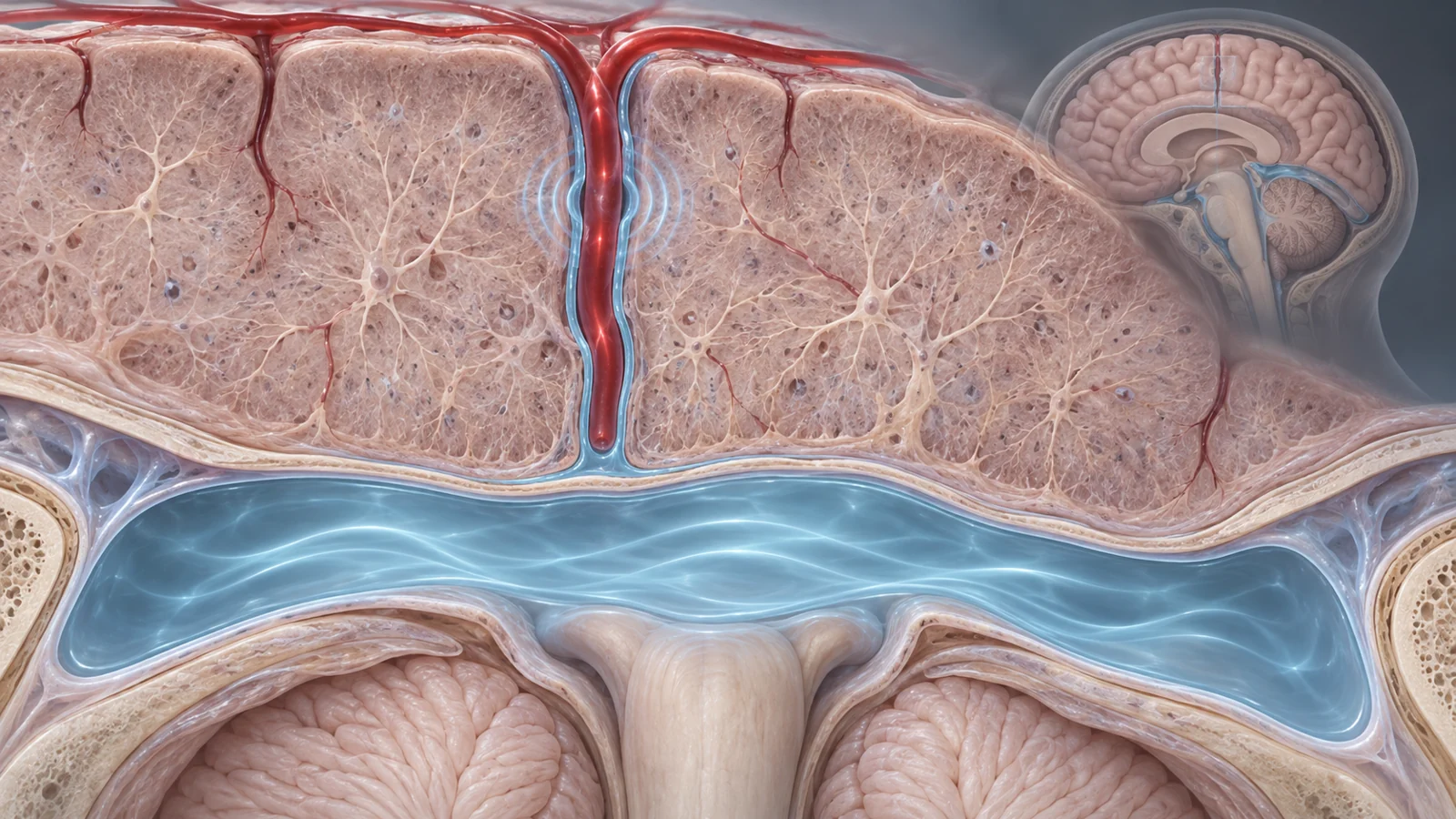
Brain fluid moves in spaces throughout and around the brain. Some of those spaces are tiny channels that run alongside blood vessels deep in brain tissue. Researchers have proposed that cerebrospinal fluid—the liquid around the brain and spinal cord—helps carry substances away from brain tissue through these routes. But seeing fluid move is not the same as measuring what it carries or how much leaves the tissue.
A study published on October 14, 2025, introduces a way to measure cerebrospinal fluid mobility in living people at a scale that reaches spaces around small penetrating vessels. The researchers used it to compare how heartbeat and breathing relate to movement in different brain regions, test whether visual stimulation changes movement locally, and explore differences in people with cerebral amyloid angiopathy, a disease involving amyloid buildup in brain blood vessels.
The results offer a closer look at fluid movement where clearance is thought to occur. They do not yet tell us whether a particular movement pattern improves clearance.
A scan designed to isolate fluid near small vessels
Most earlier human imaging of cerebrospinal fluid focused on larger spaces, such as the fluid around the brain or inside its ventricles. Those measurements could not resolve the small perivascular spaces around penetrating vessels, which are thought to connect brain tissue with larger fluid spaces. Studying those channels in living people could help clarify how movement varies across locations without injecting a tracer into the fluid.
The team developed an MRI method called CSF-STREAM. It uses the magnetic properties of cerebrospinal fluid to suppress signals from blood and brain tissue. The resulting images isolate the fluid signal, including in small perivascular spaces. Motion-sensitive gradients are applied in several directions; changes in the fluid signal are then used to calculate a measure of movement for each image volume.
The authors call this quantity “mobility” because the scan cannot distinguish among slow flow, laminar flow, back-and-forth motion, or a combination of these. In other words, it measures how much motion affects the signal in a small volume, but not whether fluid travels steadily in one direction. A mobility value is not a direct measurement of waste leaving brain tissue.
That distinction matters for interpreting the findings. Greater movement near a vessel could reflect different kinds of motion, and this method does not reveal how much amyloid, tau, or other material is carried along with it.
Heartbeat and breathing vary by location
In 11 participants, the researchers compared mobility fluctuations across phases of the cardiac and respiratory cycles. In selected larger spaces at the base of the brain—the space around the middle cerebral artery and the fourth ventricle—mobility varied more with the heartbeat than with breathing. In the smaller perivascular spaces in the basal ganglia and around penetrating vessels, the two rhythms had similar effects.
The regional pattern suggests that the forces associated with fluid movement are not uniform throughout the brain. In these measurements, heartbeat-related changes were more prominent in some large spaces, while pulse and breathing had comparable effects in the smaller channels. The study measured these rhythms’ association with mobility; it did not establish which rhythm moves more fluid out of brain tissue.
The researchers also checked whether their reconstruction process could itself create an apparent cycle. They compared the rhythm-based results with data assigned to random phases. Random ordering produced less coherent fluctuations and lower fit quality than assignment based on recorded heartbeats or breathing. This control supports the interpretation that the observed patterns tracked physiological rhythms rather than arising solely from reconstruction. It does not turn the mobility measure into a measure of net flow.
Visual stimulation produced a small local change
A separate experiment asked whether a regular visual stimulus could alter mobility near the visual cortex. Nine participants viewed a flashing checkerboard at 0.1 hertz while the team measured fluid movement. The average mobility in the targeted region increased by 1.2 percent during stimulation. In a comparison region outside the stimulated area, the average change was close to zero.
This experiment adds a controlled change to the observations of naturally varying heartbeat and breathing. The local difference is consistent with the possibility that stimulation-related changes in blood-vessel motion can affect nearby fluid mobility. The study did not show that the change cleared more material from brain tissue, or that the stimulation would improve clearance during sleep or benefit health.
The target area was defined using visual activation and then restricted to voxels containing cerebrospinal fluid. The result therefore describes a response in the fluid-containing area targeted by the scan, rather than a direct measurement of fluid movement inside brain cells or tissue. It is an early human observation from a small group, not evidence of a treatment effect.
The patient comparison complicates a simple reading
The team also compared eight people with cerebral amyloid angiopathy (CAA) with eight age- and sex-matched healthy controls. In the fluid space around the middle cerebral artery, the CAA group had 20 percent higher mobility and 10 percent lower fractional anisotropy, a measure of how strongly movement is oriented in a preferred direction.
Together, those measurements are compatible with more movement that is less directionally organized in this particular large space. Higher mobility here should not be read as evidence of better clearance. One proposed interpretation is that changes associated with CAA could make movement at the brain’s surface differ from movement through tissue. The study did not establish the cause of the group difference, and its small comparison cannot show whether this pattern predicts disease or patient outcomes.
The finding was also location-specific. In the sampled perivascular spaces around penetrating vessels in the centrum semiovale, mobility was not significantly different between groups. The patient comparison therefore did not show a uniform change across all measured spaces. Nor does a difference in the larger space establish how much amyloid or other material was removed from brain tissue.
A detailed measurement with practical limits
The technique’s resolution makes it possible to examine fluid near small vessels without an invasive tracer, but acquiring the measurements takes time. The whole-brain scan used for static images took about five and a half minutes; the full mobility acquisition took 38 minutes and 30 seconds. Two datasets in the younger cohort were excluded because of motion artifacts, and other participants were excluded for motion or inadequate cardiac recordings.
Long scans make it harder to repeat the measurement easily and increase the chance that movement will affect image quality. The small groups also mean that the rhythm and stimulation findings need confirmation in larger samples. The patient comparison is explicitly a pilot, so its regional results are a starting point for further study rather than a clinical benchmark.
The method also has limited ability to capture motion that is not synchronized to a measured external signal. Cardiac and breathing cycles could be recorded and used to sort measurements by phase. Vasomotion—the slow rhythmic change in blood-vessel diameter—does not have an equivalent external sensor in this study. The visual-stimulation experiment provides one way to test a related influence, but it does not resolve the underlying movement pattern or connect it directly to clearance.
What repeated brain-fluid scans could reveal
Because CSF-STREAM does not require a tracer injection, researchers could potentially repeat measurements in the same people during sleep studies or as a disease progresses. That could make it possible to track how fluid mobility near small vessels changes over time and compare those changes with other aspects of brain health. If shorter, repeatable scans are shown to reflect actual clearance and meaningful disease change, they might eventually help monitor progression in conditions such as CAA without tracer injections. That clinical use remains unproven.
Key Sources
- Primary source: Region-specific drivers of CSF mobility measured with MRI in humans.
DOI or Journal Page · Peer-Reviewed Paper · 2025-10-14
Keep exploring