
Evidence date · 2024-09-15 · Research note 2 of 2
1 2
See current Track status →
Since the previous research note · Earlier expectation not tested
Track continuity
Since the previous research note: How aging changes brain immune cells in female and male mice
Previous expectation: Treatments tailored to age- and sex-related brain immune changes might reduce damaging inflammation while preserving useful microglial functions.
What this study adds: The September 2024 review brings together senescence identification, iron handling and experimental glial-targeting treatments across neurodegenerative diseases. It supplies a broader treatment rationale without testing the earlier age-by-sex therapeutic forecast in the authorized evidence.
What changed in this study
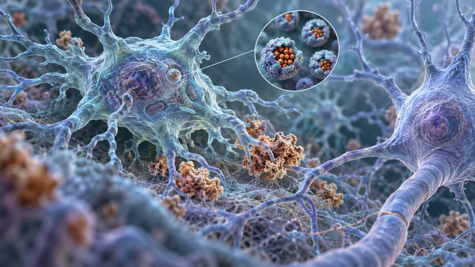
Microglia are the brain’s resident immune cells. They survey the tissue, respond to injury and infection, help maintain its environment, and remove cellular debris, including misfolded proteins. Those jobs can be protective. They can also change as the brain ages or develops neurodegenerative disease.
A review published on September 15, 2024, considers whether some disease-associated microglia enter a state called senescence. In this context, senescence means a persistent loss of the ability to divide accompanied by changes in cell behavior and secretion. Senescent cells can release inflammatory factors that alter the surrounding tissue. The possibility is important because a treatment that reduced a harmful microglial state might help the brain, but a treatment that removed useful immune cells could create a different problem.
The review brings together controlled cell experiments, observations in animal models, findings from human brain tissue, and therapeutic studies with different endpoints. Its authors reported searching PubMed and Medline in January 2024 and checking references from the selected literature. The result is a map of a promising idea, not a demonstration that selectively removing senescent microglia already benefits patients.
The brain’s caretakers can change with age
Microglia begin in the yolk sac during embryonic development and establish a self-renewing population in the central nervous system. In a healthy brain, they continually monitor their surroundings. When disease-associated proteins or damaged cells accumulate, microglia may gather around affected neurons and alter how they respond.
Alzheimer’s disease illustrates the changing balance. Early in the disease, the review describes microglia as helping to clear amyloid-beta through phagocytosis, a process in which cells engulf material. With aging and disease progression, microglia become more activated and cluster around neurons, yet their efficiency at clearing amyloid-beta can diminish. Higher inflammatory signaling may add to the damaging environment and contribute to further protein accumulation.
This pattern helps explain why “activate the microglia” or “remove the microglia” is too simple a treatment goal. The same broad cell population can perform useful cleanup in one setting and contribute to persistent inflammation in another. Researchers therefore need to identify a particular cell state, rather than treating age, activation, or an unusual shape as proof that a cell should be eliminated.
What senescence means in microglia
Senescence is not simply another word for old. It describes a cellular state involving durable cell-cycle arrest and altered activity. Senescent cells may acquire a senescence-associated secretory phenotype, or SASP, in which they release inflammatory cytokines and other factors that reshape nearby tissue.
The reviewed evidence places senescent-looking microglia in regions affected by different neurodegenerative diseases. Examples include the substantia nigra in Parkinson’s disease and the hippocampus in Alzheimer’s disease. Their location raises a plausible connection between local cellular aging and the anatomy of disease, but location alone does not establish what the cells caused or whether every cell with the same appearance shares the same state.
The label is difficult to apply because microglia can be aged, activated, dystrophic, or senescent in overlapping ways. There is no single marker that identifies every senescent cell. SASP-related inflammation may also come from non-senescent microglia responding to their surroundings. Ferritin, a protein involved in iron storage, is informative in this context, but it varies among dystrophic microglia and does not independently settle the question.
The reviewed human-brain observations make the caution concrete. Dystrophic microglia are found in aging and neurodegenerative disease, and some contain high levels of ferritin. Yet ferritin-rich dystrophic microglia were not uniformly positive for other commonly used senescence features such as SA-beta-galactosidase or lipofuscin. A visibly damaged or unusually shaped microglial cell therefore cannot simply be counted as senescent. The field’s working picture combines SASP-related changes with elevated ferritin, while recognizing that the combination still needs careful interpretation.
The clues from tau and an ALS model
One line of evidence comes from a controlled experiment using primary microglia. Researchers exposed the cells to monomeric tau at 5 or 15 nanomoles per liter for 18 hours, then allowed a 48-hour recovery period. In the higher-exposure condition, the reviewed study reported cell-cycle arrest, changes in cell morphology, and release of SASP factors. The result supports a connection between exposure to a misfolded-protein-related material and a senescence phenotype in microglia under laboratory conditions.
Those concentrations are experimental settings, not patient thresholds. They show how a particular exposure schedule affected cultured cells; they do not tell us what amount of tau would trigger the same state in a human brain. The experiment also does not establish that tau is the common initiating cause of microglial senescence across neurodegenerative diseases.
The ALS evidence has a different role. In the SOD1 G93A transgenic rat model, the review describes reduced lamin B1 expression in the lumbar spinal cord and increased p16Ink4a expression in microglia. Cultured microglia from the same model showed a large, flattened morphology, higher levels of several senescence-associated markers, and increased SASP factors. These are disease-associated observations in a living animal model and in cells derived from it, rather than a controlled tau exposure in otherwise isolated primary microglia.
Together, the studies suggest that more than one stress context can be associated with senescence features. They do not establish one causal sequence shared by Alzheimer’s, Parkinson’s, Huntington’s disease, and amyotrophic lateral sclerosis. The value of comparing them is more modest and more useful: it shows why researchers are asking whether senescence is a recurring cell state across diseases, while keeping each model’s evidence in its proper setting.
Why ferritin complicates the picture
Ferritin stores iron inside cells. For microglia, that storage can be protective for nearby neurons because it helps sequester free iron that might otherwise participate in reactions producing highly reactive oxygen species. In a diseased brain, however, the same iron-handling burden may contribute to stress within the microglia themselves.
The review describes iron accumulation as a feature associated with senescent or dystrophic microglia and notes that iron overload has been linked to senescence in laboratory studies. It also emphasizes an unresolved question: how much of the iron accumulating in brain tissue comes from local handling of disease-related material, and how much might enter through the blood-brain or blood-cerebrospinal-fluid barriers and be distributed through other pathways. The evidence supplied here does not turn that uncertainty into advice about dietary iron, blood iron, or supplements.
Ferritin is consequently a useful clue with two meanings. It can indicate an attempt to contain potentially damaging iron, while also pointing to a cell under metabolic or oxidative strain. A ferritin-rich microglial cell may be protecting neurons from free iron even as its own condition deteriorates. That tension is one reason iron storage cannot be read as a simple label for “bad” cells.
It also helps explain why marker combinations matter. A rise in inflammatory secretions may reflect senescence, but it may also reflect the broader inflammatory environment. Ferritin may identify a particular iron-handling burden, but not every ferritin-rich cell has the same complete senescence profile. Separating these possibilities is essential before a therapy is designed to remove or reprogram a population.
Two treatment ideas with different targets
The review discusses two broad therapeutic approaches. Iron chelators bind iron, aiming to alter the availability of iron that can contribute to oxidative stress. Senolytics, by contrast, are intended to induce the death of senescent cells. The distinction matters: one approach changes a chemical burden, while the other removes cells identified as part of a harmful state.
Iron chelation has produced uneven findings in the material reviewed. Deferiprone was reported to chelate iron in the substantia nigra and improve motor symptoms in some Parkinson’s disease research, while another reported result found it ineffective in newly diagnosed patients. These contrasting outcomes do not support a general conclusion that reducing brain iron will slow neurodegeneration. They show why a plausible mechanism and a clinical effect must be assessed separately.
Senolytic evidence in the review is especially notable for its attribution limits. In a mouse model of tau-dependent neurodegeneration, the drug navitoclax, also known as ABT263, reduced p16Ink4a-positive senescent astrocytes and microglia in the cortex and hippocampus. The treated mice also showed improved memory. Because both astrocytes and microglia were reduced, the result cannot identify microglial removal as the sole reason for the memory improvement.
The review also describes work with dasatinib and quercetin in mouse models and mentions investigations of dasatinib plus quercetin in people with early Alzheimer’s disease. At the September 2024 publication point, those human investigations should be understood as efforts to determine whether senolytic treatment can affect senescent cells and SASP-related effects, not as established evidence of a safe, microglia-specific treatment that improves patient outcomes.
A further possibility discussed in the review is metformin, which has been reported in experimental Alzheimer’s and Parkinson’s settings to inhibit aspects of cellular senescence. Such findings broaden the list of ways researchers might influence a senescence-related process, but they do not resolve which cell types should be changed, removed, or preserved in a patient.
What selective treatment could mean for brain function
The practical hope is not simply to clear more cells. It is to reduce a damaging glial state while retaining the brain’s ability to clear debris, respond to injury, and support neurons. If future work can identify senescent microglia reliably, target them without eliminating protective microglia or other essential cells, and show a safe benefit in humans, treatment could eventually help preserve memory or other functions affected by neurodegenerative disease.
That possibility remains conditional. The reviewed mouse result involved both microglia and astrocytes, the iron findings do not define a universal disease mechanism, and the supplied evidence does not establish patient benefit from microglia-specific senolysis. Still, the combination of cell-state identification, iron biology, and targeted intervention gives researchers a concrete route toward a more selective form of treatment—one designed to protect brain function by changing the harmful response without discarding the brain’s immune maintenance system.
Key Sources
- Primary source: Microglial senescence in neurodegeneration: Insights, implications, and therapeutic opportunities.
DOI or Journal Page · Review or Meta-Analysis · 2024-09-15