
Research Signal – Source date 2021-02-02
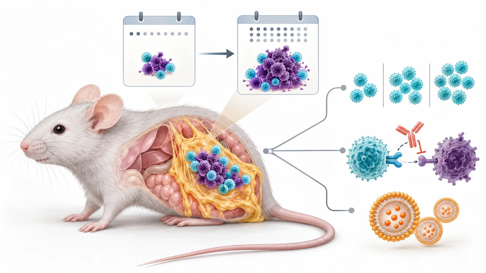
Patient-derived T cells initially slowed melanoma tumors in mice, but that early control did not last. By day 25, the tumors had grown again. The change gave researchers a way to test not only whether immune cells could recognize a tumor, but also how different cell-transfer schedules and treatments affected the later escape.
The study, published February 2, 2021, describes a model called the X-mouse. It was designed to let researchers observe human tumor-specific T cells inside a growing tumor and compare the tumor’s response over time. The experiments showed that an early drop in tumor growth could be followed by a very different outcome, even when the transferred T cells remained in the tumor.
A human tumor target in a mouse setting
The model joined two human cell populations with an immunodeficient mouse. Researchers injected melanoma cells into the abdomen of NSG mice, whose immune systems are severely limited. The tumor cells settled in the greater omentum, a small, well-supplied layer of fatty tissue between abdominal organs.
The researchers used DM6, a human melanoma cell line, as the target. They engineered these cells to display selected tumor mutations and carry green fluorescent protein, or GFP, which made them visible in tissue. Some versions also carried luciferase, a marker that produces light for imaging living mice. Together, the markers let the team measure tumor burden in the omentum after examination and follow growth and spread in the same animal by imaging.
The other human cells were CD8 T cells collected from melanoma patients who had taken part in a personalized neoantigen vaccination trial. These T cells had been selected and expanded to recognize particular mutated tumor peptides. In this experiment, the T cells came from one patient and were matched to the engineered DM6 targets. The mouse provided a controlled place to bring the two human cell types together; it did not recreate a patient’s full immune system.
Without transferred tumor-specific T cells, the melanoma cells grew rapidly and spread. With T cells recognizing a displayed mutation, tumor burden was lower at day 10. The early result demonstrated that the cells could act against a matched target in this setting. The later measurements asked a harder question: would that control endure?
Early control did not guarantee lasting control
By day 25, the matched tumors had escaped the initial T-cell control and grown substantially. In untreated mice with progressing tumors, the researchers also observed abdominal fluid and tumor deposits in other organs. Imaging in living mice showed the same broad pattern: growth was suppressed after the T-cell transfer, then began to rise again.
The team tested whether the return could be explained simply by too few T cells. Increasing the initial number of transferred cells improved early suppression, but did not prevent later escape. The degree of escape was smaller with higher cell doses, yet recurrence still occurred. T cells were also detectable in the tumors at day 25, so their disappearance from the tumor was not a sufficient explanation for the change.
The researchers examined other features of the tumor that could affect recognition. They found no evidence that the tumors had lowered the tested major histocompatibility complex class I, a surface system involved in displaying peptide targets to T cells. The tumor cells also retained strong GFP expression, used as a marker for the engineered constructs carrying the target peptides. These checks narrowed the possibilities, but did not identify a single complete cause of escape.
Instead, measurements pointed toward changes in the T cells and their surroundings. T cells isolated from tumors showed increased PD-1, an immune checkpoint receptor that can reduce T-cell activity when engaged. LAG3 and other checkpoint markers were also associated with persistent stimulation in the researchers’ experiments. The tumor cells increased PD-L1, a molecule that can engage PD-1, after exposure to material from activated T cells. These observations made checkpoint signaling a plausible treatment target to test; marker changes alone could not prove that they fully explained the escape.
Different cell schedules changed the later outcome
The researchers compared ways of broadening or refreshing the T-cell response. One approach combined two populations of T cells from the same patient, each recognizing a different tumor mutation. Both single-population groups showed early tumor suppression. By day 25, however, the combined group maintained better control, while tumors in the single-specificity groups had grown again. In this model, targeting two mutations provided more durable control than either tested specificity alone.
A separate comparison changed timing rather than target breadth. Mice received T cells recognizing one mutation either once, on day 5, or again on day 10. The second transfer significantly suppressed the later escape observed after a single transfer. These experiments suggested that both the range of tumor targets recognized and the timing of cell delivery could affect how long control lasted. They were distinct protocol choices, not evidence that one schedule would suit every tumor.
Two treatments tested different ways to support T cells
The researchers next tested nivolumab, an antibody that blocks PD-1. In this setup, PD-1 blockade prevented the tumors from escaping T-cell control by day 25. Because PD-1 had increased on the transferred cells, the result supported a role for this checkpoint in the model and showed that the X-mouse could register an effect from a checkpoint-directed treatment.
They also tested interleukin-12, or IL-12, delivered in liposomes. IL-12 is a signaling protein used here to stimulate T cells. A single treatment partially suppressed later tumor escape. When the researchers repeated the liposome treatment on days 15 and 20, the response was stronger: the schedule prevented escape and reduced tumor burden from the day-10 measurement. The treated mice also had higher circulating interferon-gamma, a marker consistent with increased T-cell stimulation.
These results should be kept separate. Nivolumab blocked a checkpoint pathway and prevented escape in the reported comparison. IL-12 provided a T-cell-stimulating treatment whose effect depended on the schedule: one dose had a partial effect, while repeated dosing produced a stronger response. Neither experiment established a treatment benefit or safe dose for people.
The model makes a narrow question easier to study
The X-mouse was built to isolate the interaction between tumor cells and transferred tumor-specific T cells. That simplifies interpretation: an observed change after an immune treatment can be studied within this pair, with fewer other immune-cell interactions to account for. Researchers could also recover cells from the omental tumors and examine their number and characteristics, alongside changes in tumor burden.
The same design narrows what the findings can show. The featured experiments used one engineered melanoma cell line, with tumor targets built to display selected mutations. Tumors from different patients can vary, and the model’s results do not establish that the same cell combinations or schedules would work across that diversity. The immunodeficient mouse also lacks much of the immune setting present in people. And although the omentum offered a convenient, consistent site for growing and measuring tumors, it is not a usual site for melanoma in patients.
The study therefore supports the model as a controlled preclinical test bed, especially for approaches intended to improve T-cell function. Its value lies in making the time course visible and allowing treatment comparisons under consistent conditions. Whether those comparisons predict responses in people remains an open question.
What a faster immune therapy test could change
If testing in varied tumors shows that this model predicts human responses, researchers could use it to choose T-cell combinations and schedules for clinical trials that aim for lasting control, rather than a short-lived initial response. For patients, the potential payoff would be treatments that keep tumors in check longer. These mouse experiments cannot show whether any of the tested approaches will do that in people.
Key Sources
- Primary source: Preclinical evaluation of cancer immune therapy using patient-derived tumor antigen-specific T cells in a novel xenograft platform.
DOI or Journal Page · Peer-Reviewed Paper · 2021-02-02
Keep exploring