
Research Signal – Source date 2026-05-20
A liver tumor is more than a collection of cancer cells. It sits among immune and other cells that can either support an attack or help the cancer evade one. That surrounding community varies between tumors, and it can shape how well immunotherapy works. A model that grows tumor tissue together with immune cells gives researchers a way to examine some of those interactions in the laboratory.
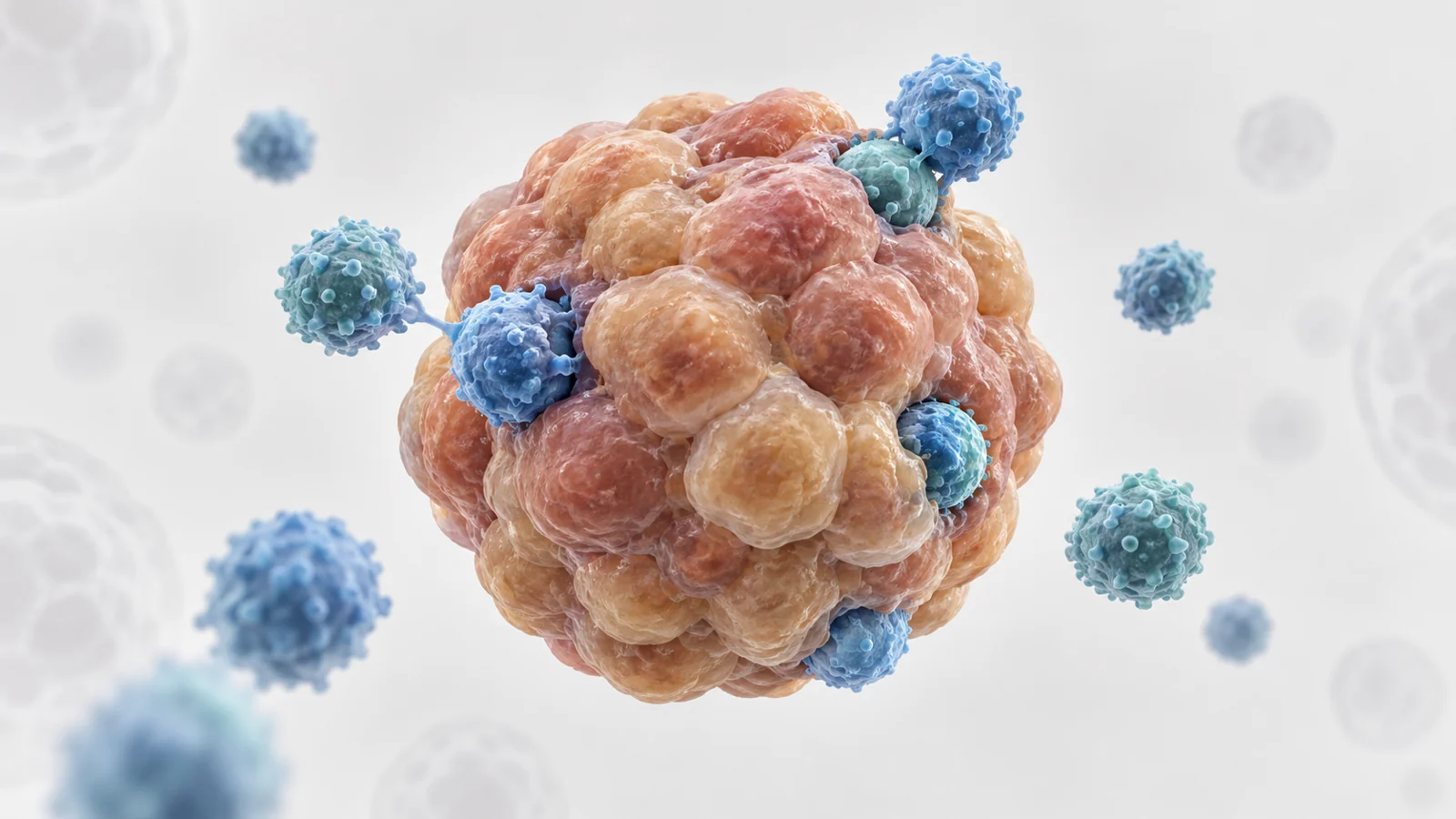
A review published on May 20, 2026, describes platforms that combine liver cancer organoids with defined immune-cell populations. An organoid is a small, three-dimensional model grown from tumor tissue that can retain some features of the original tumor. Pairing one with immune cells can help researchers investigate how immune activity changes around cancer cells, and how a treatment might alter that activity.
The approach offers a closer view of tumor–immune interaction than cancer cells grown alone. But the details of the model matter: whose immune cells are added, how the cells are arranged, and what researchers measure all affect what an experiment can say.
The immune-cell source changes the question
Researchers can establish liver cancer organoids from fresh tumor tissue collected at surgery or biopsy. The tissue is broken down and embedded in a three-dimensional matrix, with growth conditions chosen to support organoid formation. Media and matrix recipes can affect how well cultures grow and which tumor features remain represented. Conditions that support long-term expansion may also favor some cell populations over others, so a growing organoid need not capture every feature of the original tumor equally.
The immune cells added to an organoid also shape the experiment. When they come from the same patient—such as that person’s peripheral blood immune cells or tumor-infiltrating lymphocytes—the pairing can preserve more of the patient-specific context. Researchers can ask how that patient’s tumor and immune cells interact under selected conditions.
Immune cells from an unrelated donor offer a different kind of experiment. Donor cells and established cell lines can be easier to obtain or use consistently, but they do not provide the same match to the tumor or the same tumor-specific recognition. Such pairings can help study general interactions, while limiting what can be concluded about a particular patient’s immune response.
These choices set the scope of the model. A tumor organoid combined with a defined immune population can isolate a specific interaction for study; it does not recreate the full immune community found in a person’s liver tumor.
Checkpoint signals can weaken T cells
One interaction described in the review involves PD-L1 on tumor cells binding to PD-1 on T cells. The signaling can impair T-cell activity: researchers have observed reduced proliferation, lower release of inflammatory signaling molecules such as IFN-γ and TNF-α, and diminished ability to kill tumor cells. Other inhibitory receptors, including Tim-3 and Lag-3, may add to T-cell dysfunction.
In co-culture experiments, researchers can examine these changes with the tumor and immune cells present together. The review describes partial recovery of some T-cell functions after checkpoint blockade, such as treatment targeting PD-1. In this setting, a recovery suggests that the pathway can contribute to suppression under the tested conditions. It does not show that every source of immune dysfunction has been reversed, or that the same response will occur in a patient.
The model can also be used to investigate other interactions. The review discusses adenosine-related metabolic suppression and chemokine-regulated immune recruitment among the processes examined in co-culture systems. Together, such experiments can help researchers probe how tumors create a less favorable setting for immune attack and how immune cells respond when parts of that setting are altered.
Contact and flow shape what can be observed
In direct co-culture, immune cells and organoids share the same environment. Immune cells can contact and enter the tumor-like structures, allowing researchers to observe interactions and measure cell killing. That close contact is useful for studying cytotoxicity, though it can also produce overactivation, rapid exhaustion, or uneven expansion of cell populations.
Indirect arrangements keep the cell populations apart while allowing some communication between them. This can help researchers focus on signals exchanged between tumor and immune cells without direct contact. The setup cannot answer the same questions about immune-cell entry or direct killing as a shared environment.
Static three-dimensional cultures, often grown in multiwell plates, are relatively simple and can preserve organoid structure. They are useful for early screening and focused experiments. But diffusion is limited, and keeping immune cells viable over longer periods can be difficult.
Dynamic platforms, including microfluidic organ-on-chip systems, allow researchers to study interactions as conditions change over space and time. Some can model features such as low-oxygen regions and the gradients associated with them. The review describes how hypoxia can be linked to changes including PD-L1 and CD73 expression, which may contribute to an adenosine-rich, immunosuppressive environment. Such systems offer ways to explore these relationships under controlled conditions, but they also add complexity to the design and interpretation of experiments.
No single configuration answers every question. A simple static culture may suit a screening task, while dynamic systems can be useful when movement, gradients or changing conditions are central to the biological question.
Killing tumor cells is only one measure
Researchers can measure tumor-cell killing with assays that track cell viability or damage, including ATP-based viability, lactate dehydrogenase release and real-time impedance. Imaging can follow changes in organoid growth. These measurements address whether tumor cells are damaged or reduced under the conditions of the experiment.
Immune-cell function calls for separate readouts. Researchers can measure activation markers such as CD69 and CD25, effector molecules such as IFN-γ and granzyme B, and inhibitory receptors associated with exhaustion, including PD-1, Tim-3 and Lag-3. Looking at these alongside tumor-cell measurements helps distinguish a strong short-term killing result from evidence that immune cells remain active during sustained exposure.
That difference matters because primary immune cells may not persist well in culture, and their phenotype can shift under laboratory conditions. A brief experiment may capture an initial burst of cytotoxicity while missing later exhaustion, functional adaptation or changes in the tumor cells selected by immune pressure. Duration and cell state therefore influence what a killing measurement can mean.
Missing tissue context leaves gaps
Most co-cultures simplify the tumor environment. Many do not include the supporting stromal cells, functional blood-vessel lining or perfusable vessels present in living tissue. Without these features, immune-cell movement into a tumor-like structure, the supply of oxygen and nutrients, and the distribution of a treatment may differ from what happens in a person.
Laboratory methods add another source of variation. Matrix composition, growth medium, the ratio of immune cells to organoid cells and the choice of measurement can all differ between studies. Matrices can vary from batch to batch, and researchers may define a treatment response differently depending on the assay. These differences make results harder to compare across laboratories and patients.
The practical requirements also matter. Maintaining organoid collections and operating advanced co-culture platforms can require specialized infrastructure and resources, limiting how widely the approaches are available.
Where patient-matched tumor tests could lead
Patient-derived organoids paired with a patient’s own immune cells could eventually help researchers compare candidate immunotherapies or combinations in a setting that reflects some features of that person’s tumor. If standardized assays show that these results reliably track treatment outcomes, the approach could support more individualized treatment research and help identify promising strategies before they are tested in patients. For now, the review describes potential rather than a clinically established selection tool: prospective studies linking model results to patient outcomes remain necessary, particularly given the influence of liver disease and differences among patients.
Key Sources
- Primary source: A review of organoid-immune co-culture platforms to model the immune microenvironment of hepatocellular carcinoma and guide immunotherapy.
DOI or Journal Page · Review or Meta-Analysis · 2026-05-20
Keep exploring