
Research Signal – Source date 2025-12-22
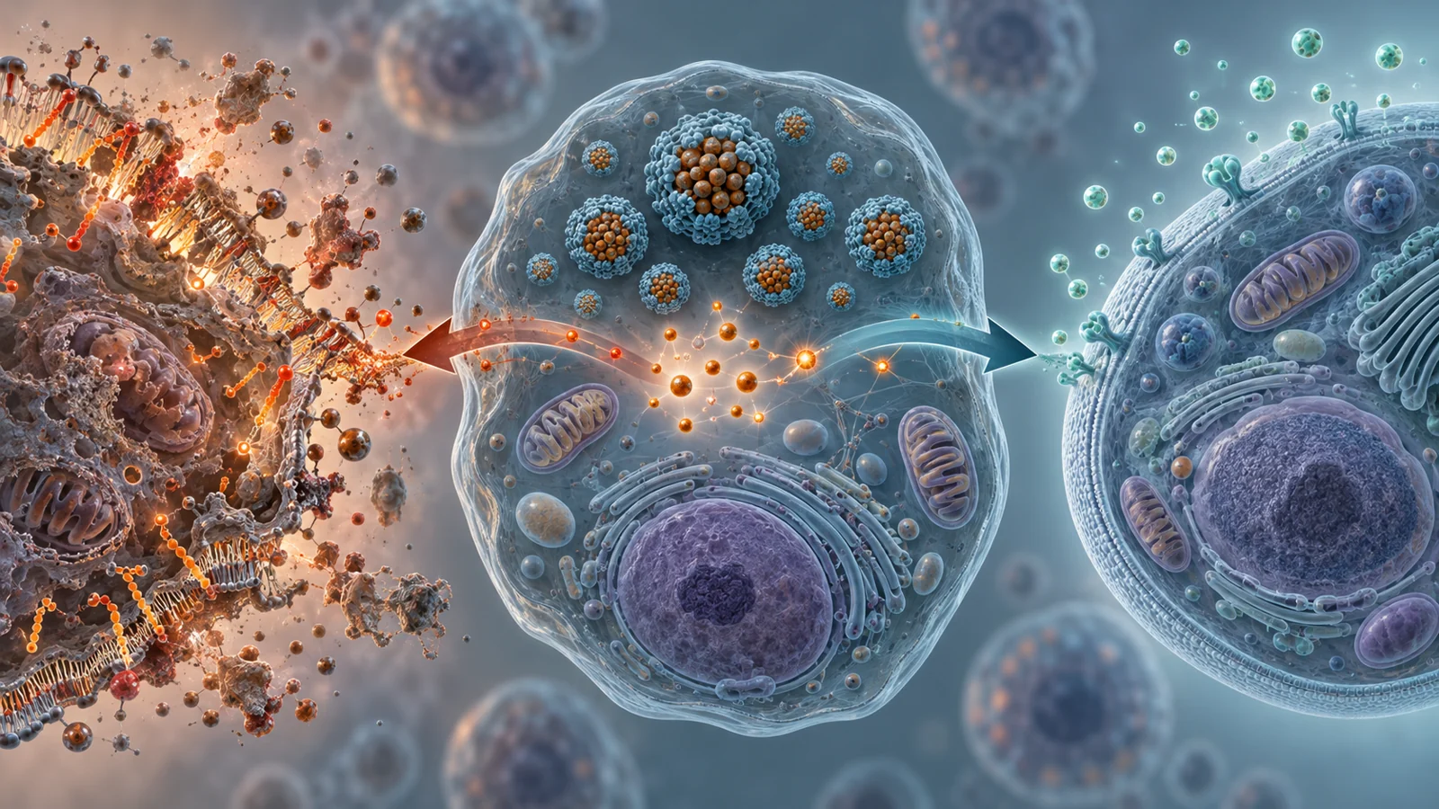
A review published on December 22, 2025, brings two cellular processes into the same conversation: ferroptosis, an iron-dependent form of cell death driven by lipid damage, and senescence, a persistent state in which a cell stops dividing. Both can be shaped by iron and oxidative stress, but they are not the same event and one is not simply a delayed version of the other.
That difference matters because a treatment designed to protect cells in one disease setting might be useful precisely because it kills cells in another. The practical question is not whether iron-related chemistry is always harmful. It is which cell is affected, what state that cell enters, and whether changing that state helps the surrounding tissue.
The iron inside a cell is not all equally reactive
Cells need iron for essential tasks, including the production and maintenance of iron-containing proteins. Blood transports iron through transferrin, which delivers it to cells through transferrin receptors. Once inside, iron can enter a low-molecular-weight transit pool or be stored in ferritin. Storage and controlled use help keep the metal available for biology without allowing it to drive uncontrolled chemical reactions.
The chemically available portion is more important to oxidative injury than the total amount of iron measured in a cell. When the labile iron pool expands, iron can participate in reactions that generate highly reactive molecules. Those molecules can damage DNA, proteins, mitochondria, and the fatty components of cell membranes. In senescent cells, altered iron export and impaired ferritinophagy may contribute to iron accumulation, helping maintain a redox-imbalanced state and the inflammatory secretions associated with senescence.
Ferroptosis adds another layer. The cell membrane contains polyunsaturated fatty acids that can be incorporated into phospholipids. Enzymes such as ACSL4 help establish a lipid pool that is especially vulnerable to oxidation, while lipoxygenases and related enzymes can promote the formation of lipid peroxides. If those peroxides accumulate faster than the cell can neutralize them, the membrane reaches a damaging threshold and ferroptotic death follows.
One of the main protective systems involves glutathione and GPX4. GPX4 helps convert lipid hydroperoxides into less damaging products, using glutathione as a cofactor. Limiting cysteine import, depleting glutathione, or directly inhibiting GPX4 can weaken this defense. Other pathways, including FSP1 with coenzyme Q10 and NAD(P)H, provide additional protection in membranes, while GCH1-BH4 and DHODH help protect particular cellular compartments.
The review therefore connects the two processes through a shared chemical environment rather than a shared fate. Persistent, sublethal oxidative stress can help push a cell into stable growth arrest, with a secretory profile that affects neighboring cells. A more acute failure of lipid-peroxide control can instead produce ferroptosis. The timing, intensity, and cellular response alter the biological result.
The desired outcome can reverse from one cell type to another
Liver disease shows why a single pathway cannot be labeled simply good or bad. In fibrosis and cirrhosis, ferroptosis of hepatocytes can promote fibrogenesis and worsen the damage that supports scar formation. Yet the review describes experimental evidence suggesting that selectively inducing ferroptosis in hepatic stellate cells could reduce matrix deposition. The same broad form of cell death could therefore be damaging in the cells that maintain liver function and potentially useful in cells that help build excess scar tissue.
A similar logic appears in cancer research. Some treatment-resistant tumors rely heavily on GPX4 or alternative antioxidant systems such as FSP1-coenzyme Q10. In experimental models, disrupting those defenses or otherwise inducing ferroptosis has been investigated as a way to resensitize cancer cells to chemotherapy, radiotherapy, or targeted inhibitors. Ferroptotic cells may also release danger signals that support antitumor immune responses.
The risk is that ferroptosis is not automatically tumor-selective. Lipid-peroxidation products released during chronic injury may contribute to inflammation and, under some conditions, support tumor-promoting processes. The challenge is to exploit a weakness in established tumor cells without creating the same oxidative injury in healthy tissue. The liver example makes the principle concrete: the therapeutic goal depends on the identity and role of the cell, not just on the name of the pathway.
Senescence calls for a different set of choices. A senescent cell remains alive but has permanently exited the cell cycle. DNA-damage responses involving p53 and p21 or p16 and Rb help enforce that arrest. Mitochondrial dysfunction can raise reactive oxygen species, while changes to chromatin and loss of lamin B1 help stabilize the state. Senescent cells can also release a senescence-associated secretory phenotype, or SASP, that promotes inflammation and tissue remodeling.
Removing those cells is not the same as quieting their secretions. Senolytics aim to eliminate senescent cells. Senomorphics aim to suppress harmful features such as the SASP while leaving the cells in place. Iron chelation is a third kind of intervention: chelators bind iron and can reduce its reactive availability. These categories may overlap in a drug’s effects, but they describe different therapeutic actions and should not be treated as interchangeable labels for anti-aging treatment.
Proposed interventions range from iron control to cell removal
Iron chelators such as deferiprone, deferoxamine, and deferasirox have established roles in managing iron overload. The review discusses their possible use against ferroptosis and senescence because reducing labile iron could lower iron-catalyzed oxidative injury. In experimental models, chelation has inhibited ferroptosis in diseases involving cancer, neurodegeneration, kidney injury, cardiac injury, and other organ damage. In senescence-related work, chelators have shown senomorphic or senolytic effects in settings including liver fibrosis and osteoarthritis models.
Those observations do not turn iron chelators into general treatments for aging. Iron is also necessary for normal cellular functions, and excessive chelation can create its own problems. The review describes variable results across diseases and emphasizes practical constraints such as route of administration, cumulative toxicity, and the need to monitor organs including the kidneys and liver. Deferiprone’s ability to cross the blood-brain barrier has made it relevant to some neurological investigations, but outcomes vary with disease context and dosing. Deferasirox has been studied in aged animal models, yet its safety monitoring remains important.
Other approaches act on the survival systems of senescent cells. Dasatinib plus quercetin has been explored as a senolytic combination, while navitoclax has shown senescent-cell clearance in preclinical models. Rapalogs and nutraceuticals such as quercetin have also been discussed as senomorphic approaches. The fact that a compound can affect iron binding, antioxidant chemistry, or a stress pathway does not by itself establish that the compound will selectively remove harmful senescent cells in people.
The therapeutic direction can also be deliberately opposite in cancer. Sorafenib and conventional drugs such as cisplatin and doxorubicin are discussed in connection with ferroptotic mechanisms, including changes in iron handling, glutathione metabolism, and lipid peroxidation. These mechanisms may contribute to anticancer activity, but they can also contribute to harm in healthy organs. The review highlights doxorubicin-associated cardiotoxicity as an example of why triggering oxidative cell death in a tumor while protecting cardiomyocytes is a difficult selectivity problem.
What the early human findings actually establish
The most useful human example in the review is dasatinib plus quercetin, because it shows both the promise and the narrowness of early translation. Studies summarized for idiopathic pulmonary fibrosis and Alzheimer’s disease reported feasibility and short-term tolerability. In the Alzheimer’s phase 1 study, dasatinib reached the central nervous system and senescence-related cerebrospinal-fluid biomarkers were modulated. In pulmonary fibrosis, participants showed modest improvements in physical function and reductions in circulating senescence markers after dosing. Exploratory work in diabetic kidney disease reported lower p16INK4a and p21CIP1 expression in peripheral blood cells, consistent with target engagement.
These are different observations, not one common measure of clinical success. Feasibility asks whether a treatment can be administered and studied. Tolerability describes how participants handled it over the observed period. Biomarker changes can indicate that a biological pathway was affected. A modest functional observation is closer to a patient-relevant outcome, but it still does not establish durable improvement in disease or an extension of life.
The review reports that adverse events in these studies were mostly mild and transient, including fatigue, gastrointestinal upset, and brief cytopenias, while also noting that long-term data remain limited. It reports no consistent changes in systemic iron indices in the dasatinib-quercetin trials. Quercetin has iron-binding and antioxidant properties, but those biochemical features do not show that the combination works by changing whole-body iron status or that its effects depend on iron.
Navitoclax supplies a different warning. Its ability to inhibit BCL-2 family proteins can clear senescent cells in preclinical models, but BCL-xL inhibition in platelets produces predictable, dose-dependent thrombocytopenia. Intermittent dosing and more selective inhibitors have been investigated to address that constraint. Biological activity and a usable treatment are therefore separate questions: a drug may hit the intended cell-survival pathway and still have a safety profile that limits how it can be used.
The same caution applies to the review’s broader discussion of proposed ferroptosis treatments. Iron chelators, antioxidants, repurposed drugs, and combinations may be useful in selected settings, but the supplied studies span different diseases, cell types, endpoints, and stages of investigation. They do not establish that lowering iron generally slows human aging, nor do they provide a common estimate of benefit across neurodegeneration, cancer, fibrosis, and organ injury.
What more selective treatments could make possible
For patients, the most consequential future possibility is a treatment that recognizes which cells are causing damage and changes their fate without disrupting useful tissue. In cancer, that could mean making a resistant tumor more vulnerable to ferroptosis while limiting the oxidative injury that can contribute to heart toxicity from treatment. In fibrotic disease, it could mean protecting hepatocytes while selectively removing or disabling cells that drive excess matrix deposition. In senescence-associated disease, it could mean eliminating harmful cells or quieting their secretions without broadly disturbing normal repair.
Those benefits remain conditional on achieving cell selectivity and demonstrating a favorable clinical balance between benefit and harm. The review’s research direction supports the possibility of more precise combinations of iron control, antioxidant defense, lipid-peroxide regulation, and senescent-cell targeting; it does not establish that this precision has already been achieved. If it can be achieved, patients could gain disease control with less collateral organ injury than comes from treating a shared chemical pathway indiscriminately.
Key Sources
- Primary source: Targeting Aging and Diseases Associated with Ferroptosis and Senescence Through Modulation of Iron, Oxidative Stress and Lipid Peroxidation.
DOI or Journal Page · Review or Meta-Analysis · 2025-12-22
Keep exploring