
Research Signal – Source date 2024-10-11
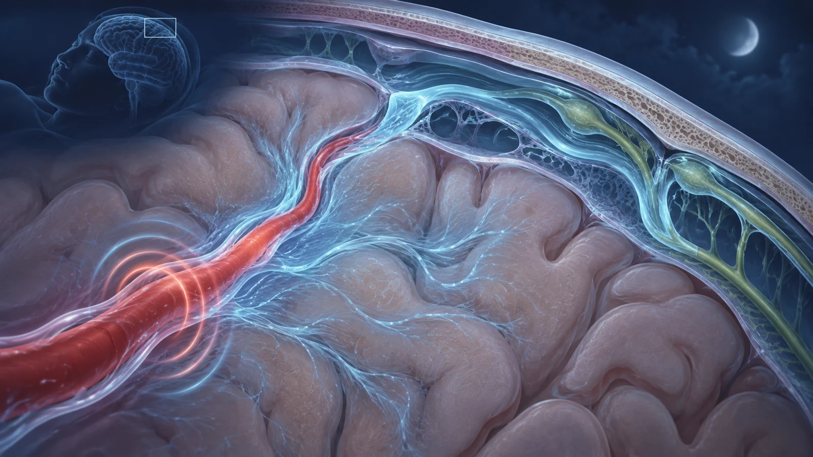
The brain relies on moving fluid to carry away some of the by-products produced by its activity. A 2024 review in Neuron describes this clearance as a connected route: fluid travels alongside arteries, passes through brain tissue, and drains toward lymphatic vessels in the membranes around the brain. Blood-vessel motion, immune cells beside vessels, sleep-related brain rhythms, and lymphatic drainage all help regulate the route.
The review brings together animal experiments, human imaging and tissue observations, and studies of Alzheimer’s and Parkinson’s disease. Across these lines of work, aging is associated with changes that could make fluid movement and drainage less efficient. The findings offer plausible ways to investigate dementia risk, but they do not show that restoring clearance prevents either disease in people.
A route shaped by vessels and the spaces around them
Fluid moves along spaces around arteries before entering brain tissue and eventually reaching drainage pathways around the brain. Arterial pulsations and local changes in blood pressure help drive the movement. The route depends on the spaces around vessels remaining open enough for cerebrospinal fluid, or CSF, to pass.
Blood flow is also adjusted moment by moment to meet the energy demands of active brain regions. This process, called neurovascular coupling, involves neurons, astrocytes, and cells in and around blood vessels. The review describes evidence that these coordinated vascular responses also help transport CSF. A change in vessel behavior may therefore affect both the delivery of blood and the movement of fluid through nearby spaces.
In rodents and humans, neurovascular coupling declines with age. The review points to several age-associated vascular changes, including increased arterial twisting and stiffness, changes in resting vessel diameter, and altered blood-brain barrier permeability. In aged mice, imaging localized impaired responses mainly to the precapillary sphincter, where small arteries transition toward the capillary bed. The precise cellular and molecular causes remain incompletely understood, but reduced vascular responsiveness could disrupt the pulsations that help move fluid.
The immune cells that help keep the route open
Perivascular border macrophages sit in fluid-filled spaces around brain vessels other than capillaries. They help manage the extracellular matrix, the material surrounding cells that can accumulate along vessel walls. In mice depleted of these macrophages, matrix built up around vessels; the added stiffness impaired vasomotion, which helps drive CSF flow. The result gives a mechanistic example of how a local change could obstruct a larger route.
A separate body of evidence concerns aging. Studies of aged mice found that these macrophages shift toward a more inflammatory, antigen-presenting profile, with fewer cells showing markers associated with scavenging. They were also less able to engulf particles in experiments. In brain tissue from older people, researchers observed a similar shift in macrophage markers compared with tissue from younger adults. Those human tissue findings are observations; they do not demonstrate that the same functional changes directly caused impaired clearance in those people.
The review also describes evidence that macrophages interact with perivascular fibroblasts, which produce matrix. Together, the findings suggest a possible cycle: changes in immune-cell activity may allow matrix to accumulate, and a stiffer vessel environment may then make fluid movement harder. Much of the direct evidence connecting these steps comes from animal work.
Sleep rhythms and the movement of fluid
In animal studies, sleep increases CSF entry into spaces around vessels and its movement into brain tissue. Researchers have also found that sleep facilitates amyloid-beta clearance in animals. Human studies cited by the review report that a night of sleep deprivation is associated with higher amyloid-beta concentrations in CSF and reduced molecular clearance. These findings connect sleep to fluid dynamics, though they do not establish that improving sleep prevents dementia.
Brain activity during sleep changes with age. Deep-sleep slow waves and other synchronized rhythms become weaker, while older brains show less coordinated responses after sleep deprivation. Human MRI studies have measured close coupling during sleep between slow-wave activity, blood volume, and CSF pulsation, with neural activity leading the changes. This coupling offers a possible link between altered sleep rhythms and vascular motion. The human measurements do not by themselves establish that age-related rhythm changes cause a decline in waste removal.
Drainage vessels change in different ways
After fluid and its contents move through the brain, meningeal lymphatic vessels help drain CSF toward lymph nodes in the neck. With age, the initial lymphatic vessels in the meninges tend to become narrower, cover less area, and branch less. Their permeability also changes, and the downstream cervical lymph nodes can atrophy.
Some collecting-like lymphatic vessels at the base of the brain show the opposite structural pattern: they become larger and more branched. The review suggests this may be a compensatory response to weaker drainage elsewhere. At the same time, aged vessels have abnormalities in the valves that help maintain directional flow. Human MRI studies cited in the review also indicate that CSF drainage declines with age.
Because these stages connect, a bottleneck at the vessel spaces, through brain tissue, or at the final drainage route could affect the overall movement of fluid. The review describes that connected system; it does not establish that one failing stage explains age-related disease on its own.
Shared clues in Alzheimer’s and Parkinson’s disease
Alzheimer’s disease is associated with amyloid-beta plaques and tau tangles, and the review brings together findings on sleep disruption, altered CSF flow, vascular dysfunction, and changes in perivascular macrophages. Some studies link sleep patterns with tau and amyloid measures; others report impaired fluid movement in animal models and people with Alzheimer’s. These observations make clearance relevant to disease research, while leaving open whether impaired clearance is a cause, consequence, or one part of a more complex process.
For Parkinson’s disease, the review describes impaired CSF flow and lymphatic drainage, alongside sleep disorders and vascular changes. It also discusses immune activity around vessels and the recruitment of T cells in animal models and observations in patient tissue. The overlap with Alzheimer’s—sleep, vascular function, immune activity, and drainage—suggests that fluid regulation may be relevant to more than one neurodegenerative condition. It does not establish impaired clearance as the sole cause of either disease.
From promising interventions to clinical benefit
The intervention evidence addresses different steps in the chain and comes from different settings. In human studies, mild electrical stimulation and timed auditory stimulation strengthened sleep-related oscillations and improved memory consolidation measures. Those results concern sleep rhythms and memory; they do not show that the interventions increased waste removal or lowered dementia risk.
Other approaches in the review target vessels, lymphatics, or macrophages in animals. VEGF-C treatment improved meningeal lymphatic function and drainage in aged animal models. M-CSF treatment in aged animals increased matrix-degrading activity, reduced matrix accumulation, and restored CSF flow and waste removal. The review also discusses VEGF-A in relation to age-related blood-vessel changes. These experiments show that parts of the system can be altered in animals, not that the treatments are clinically effective or safe for older people.
Could restoring brain drainage help preserve memory?
If researchers can show that safely improving fluid movement in older people also protects brain function, therapies aimed at sleep rhythms, vessel motion, or lymphatic drainage could eventually help preserve memory and reduce some contributors to cognitive decline. The route from changing a mechanism to a meaningful clinical benefit remains to be demonstrated: human studies would need to connect the intervention to better clearance and then to durable cognitive outcomes.
Key Sources
- Primary source: Waste clearance shapes aging brain health.
DOI or Journal Page · Review or Meta-Analysis · 2024-10-11
Keep exploring