
Research Signal – Source date 2026-01-31
A lower brain creatine reading after hard exercise sounds like evidence that the brain spent an energy reserve. But the measurement in a new human study cannot tell that story on its own. It captured a combined chemical pool whose magnetic-resonance signal can shift for more than one reason.
The same experiment found a temporary rise in a combined glutamate-and-glutamine reading, plus a possible increase in lactate in a handful of participants. These changes offer a close look at the brain’s response during early recovery from strenuous cycling. They do not show that participants thought better, or that a single ride produced a lasting brain benefit.
A surprising change after a short, hard ride
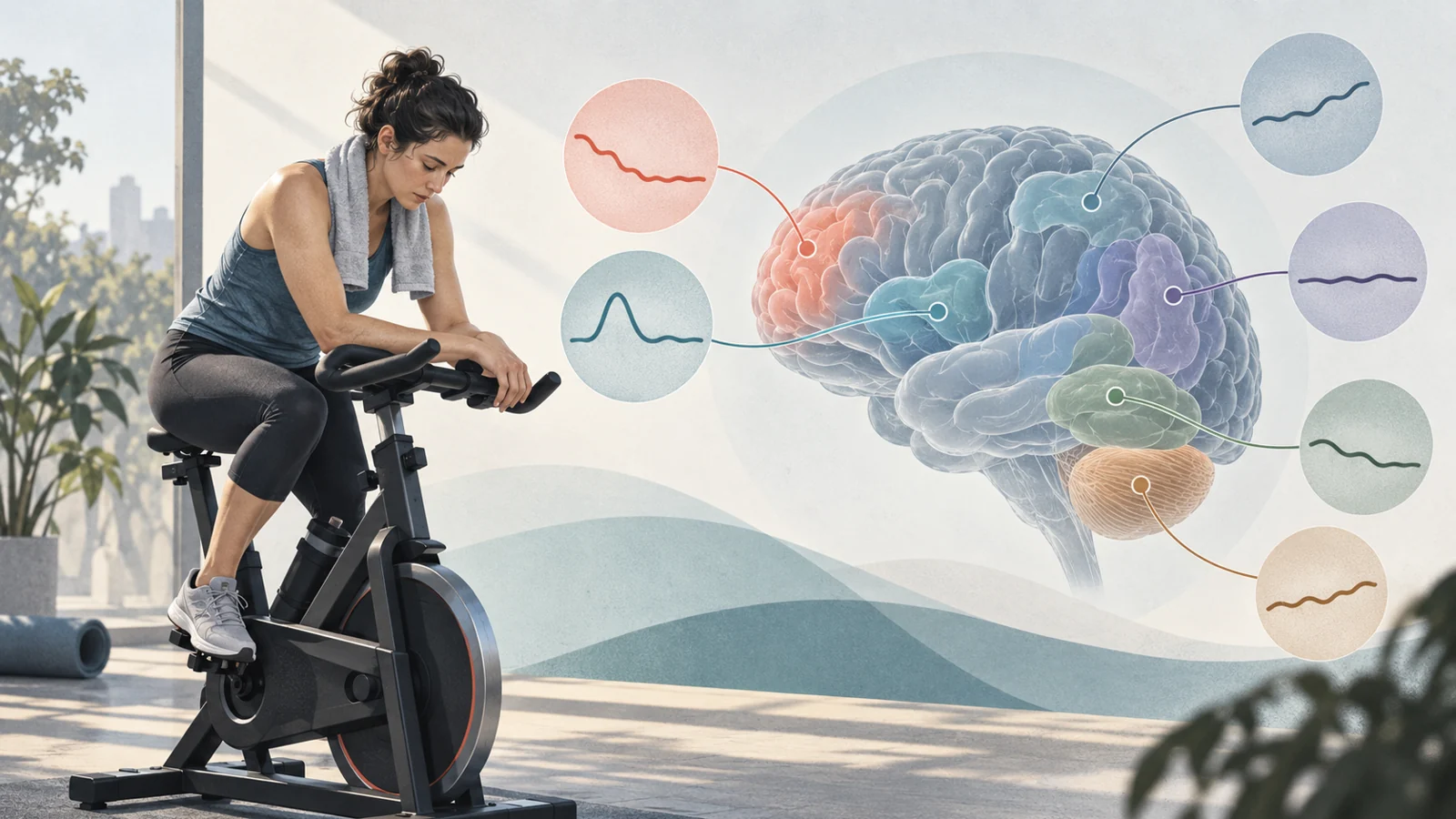
Sixteen healthy, recreationally active adults, nine of them women, completed about 15 minutes of strenuous cycling. The researchers used proton magnetic resonance spectroscopy, a method that estimates the amounts of certain chemicals in brain tissue, to scan participants before exercise and during recovery. The group was young, with an average age of 25.
The study’s planned focus was total creatine, or tCr. Creatine helps buffer energy in cells, and researchers often treat its brain level as stable enough to use as a reference for other measurements. The authors expected it to remain unchanged after exercise. Instead, in the frontal cortex, its measured concentration fell by 5.9% at 15 minutes after cycling, from 6.87 to 6.47 millimoles per liter. It was still 3.2% below the pre-exercise reading at 30 minutes, at 6.66 millimoles per liter.
The frontal cortex reading had begun to move back toward its starting level by 30 minutes. That short window leaves open what happened later, as well as what was happening during the ride itself.
A chemical reading is not a direct energy tally
At 3 tesla, the study’s spectroscopy could not separate free creatine from phosphocreatine, a form involved in cellular energy exchange. The reported tCr value combined both. If phosphocreatine is used and free creatine rises by a matching amount, the combined pool could stay steady. A lower tCr reading therefore poses a puzzle: it does not directly reveal how either form changed.
The researchers suggest that exercise may alter the local molecular environment in ways that change how visible creatine is to the scanner. Creatine and phosphocreatine have different magnetic-resonance properties; changes in their balance or interactions might affect the measured signal even if the amount of creatine in the tissue did not fall. This remains a proposed explanation, not a demonstrated mechanism.
The team checked whether a broader creatine peak might account for the change. Peak width shifted slightly at 15 minutes, but accounting for that shift still left a statistically significant difference between the pre-exercise and 15-minute readings. The checks support the reliability of the measured change; they do not settle whether the total brain creatine pool was depleted.
The pattern depended on where researchers looked
The frontal cortex was not a stand-in for the whole brain. In the visual cortex, the researchers found no reported time-related change in either tCr or Glx. A multivoxel scan, which sampled many locations across a slice of brain, found an overall 5.1% lower tCr signal and an 11.4% higher Glx signal at about 25 minutes after exercise. But the map varied by location: captured areas of the motor and sensory cortices showed the opposite directions, with tCr higher and Glx lower.
These local differences matter when interpreting an overall average. A single summary can hide areas moving in opposing directions, and this study’s results point to a spatially varied response rather than one uniform change across brain regions.
Glx rose briefly, but it combines two chemicals
Glutamate and glutamine are closely linked in the cycle through which the brain handles its main excitatory neurotransmitter. Because their signals are difficult to distinguish at 3 tesla, the researchers measured them together as Glx. This combined readout reflects aspects of glutamate and glutamine metabolism; it is not a direct count of synaptic activity.
In the frontal cortex, Glx rose 28.9% at 15 minutes after cycling, from 6.82 to 8.79 millimoles per liter. At 30 minutes it was close to the pre-exercise value, and the difference was not statistically significant. Glx was added as a research question after data collection, so this result is informative but was not the study’s original primary focus.
The rise is consistent with a temporary shift in the brain’s glutamate-and-glutamine chemistry during recovery. It cannot show that participants learned, remembered, or performed a thinking task better. Those outcomes were not measured here.
Lactate offers a limited clue
Lactate can be used in brain energy metabolism, making it a possible part of the response to strenuous exercise. In this study, however, usable frontal-cortex lactate readings were available for only five participants. In those scans, the measured level rose 76% at 15 minutes and remained 46% above baseline at 30 minutes.
The researchers treated this as exploratory. The scans were not optimized for lactate, and the small subset did not receive a statistical test. The increase may fit a hypothesis that lactate helps support energy needs during recovery, but it is not a tested result across all 16 participants.
What the measurements leave open
All post-exercise scans were taken after cycling, not during it. The final measurement came at 30 minutes, so the study does not trace the full recovery period. Nor did the researchers use phosphorus spectroscopy to measure phosphocreatine directly, which would have helped address how its level changed separately from free creatine.
The team applied spectral quality thresholds and checked how closely brain regions were repositioned between scans. Average overlap was at least 90%, and the creatine and Glx measurements met the reported quality criteria. These safeguards make the comparisons more dependable, but they cannot answer questions the measurements were not designed to resolve—especially whether the combined creatine pool truly fell or how these short-term changes relate to cognition.
The findings apply to one strenuous cycling bout in healthy, recreationally active young adults. They leave open whether the same pattern occurs in older people, people with neurological conditions, or under different exercise routines.
Why mapping the brain’s exercise response matters
A clearer account of how the brain manages energy during exercise could eventually help researchers understand which biological changes accompany exercise-related effects on thinking and brain health. If later studies link these short-term shifts to measured cognitive benefits and show which exercise routines reliably produce those benefits, the readings could help guide exercise programs aimed at supporting cognitive health. This study offers an early measurement of those responses, not evidence that one hard ride improves cognition or prevents disease.
Key Sources
- Primary source: Strenuous Exercise Alters Brain Creatine and Glutamate/Glutamine (Glx) in Humans: Evidence From Dynamic 1 H-MRS and 1 H-MRSI.
DOI or Journal Page · Peer-Reviewed Paper · 2026-01-31
Keep exploring