
Research Signal – Source date 2023-04-20
Could helping the brain remove accumulated proteins become part of an Alzheimer’s treatment strategy? A 2023 review explores that possibility through the glymphatic system, a fluid-based waste-clearance route in the brain, and the meningeal lymphatic vessels that help drain material toward the neck.
The review does not present a new human treatment trial. It brings together animal experiments, mechanistic studies, human imaging, postmortem observations and clinical context. Exercise is one of its clearest examples: in young mice genetically modeling Alzheimer’s disease, running was associated with changes in fluid movement, aquaporin-4 organization, amyloid accumulation and learning. But the same exercise protocol did not produce the reported improvements in older mice. That difference is central to the scientific question. A clearance pathway may be relevant to disease without responding uniformly across ages, models or stages.
A clearance route, not a proven treatment
Alzheimer’s disease is marked by the accumulation of amyloid-beta and tau proteins alongside progressive neurodegeneration. Those features are well established, but the processes that allow abnormal proteins to build up and contribute to disease progression remain incompletely understood.
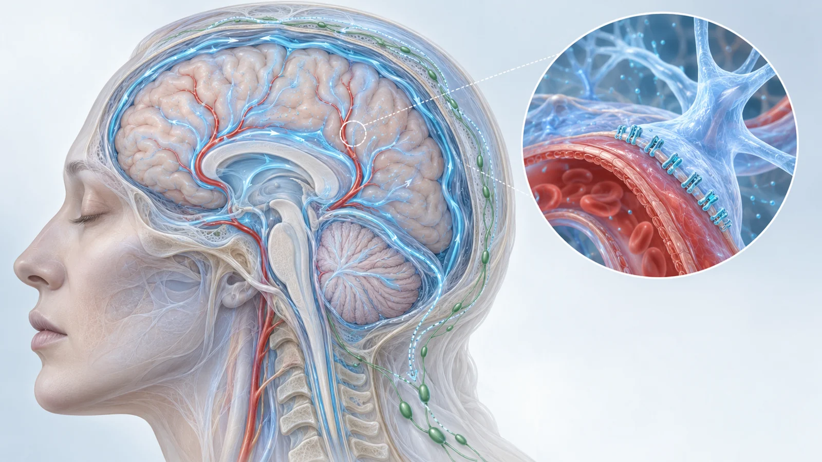
The glymphatic and meningeal lymphatic systems offer one possible part of that explanation. In the review’s account, cerebrospinal fluid moves alongside arteries, exchanges with fluid in the brain tissue, and helps carry metabolic waste toward spaces around veins. The meningeal lymphatic system is a related but distinct drainage network in the membranes surrounding the brain. Material cleared from the brain can reach these lymphatic vessels and then the deep cervical lymph nodes.
This makes the two systems interesting as connected therapeutic targets. It also creates a risk of oversimplification. Exercise, nutrition and brain stimulation affect many biological processes at once. The review therefore treats clearance as a possible contributor to their effects, not as a complete explanation for everything that happens after an intervention.
How the brain’s fluid system is supposed to work
The proposed route begins outside the brain tissue. Cerebrospinal fluid enters spaces surrounding penetrating arteries, sometimes called periarterial or Virchow-Robin spaces. From there, it moves into the brain’s parenchyma, where it mixes with interstitial fluid and travels toward perivenous spaces. The exchange can carry dissolved material through the tissue before it reaches drainage routes outside the brain.
Aquaporin-4, or AQP4, is important in this discussion because it is a water channel expressed by astrocytes and other cells. The relevant feature is not simply how much AQP4 exists. The review describes a high concentration of AQP4 at astrocyte endfeet around blood vessels. This positioning is called polarization, and the review describes it as especially important for glymphatic transport.
Aging and Alzheimer’s-related pathology can disturb that organization. In human postmortem tissue, people with Alzheimer’s disease showed increased AQP4 immunoreactivity but less polarized AQP4 than age-matched controls. Vascular amyloid deposition was also associated with lower perivascular AQP4 expression. These observations connect the location of the channel with the proposed clearance process, although they do not by themselves show that restoring its position would improve cognition.
The lymphatic side of the pathway matters as well. Tracer studies in animals have found material in basal meningeal lymphatics and cervical lymph nodes. Disrupting dorsal meningeal lymphatics can impair tracer drainage and, in some experiments, reduce glymphatic influx even without an observable change in polarized AQP4. The findings suggest functional cooperation between the two systems rather than two completely independent routes.
The underlying transport mechanism is not settled in every detail. Some experiments have emphasized convective movement, while others have reported results more consistent with diffusion or dispersion, including differences based on tracer size. A multicenter comparison of several AQP4-deficient mouse lines supported a role for AQP4 in solute uptake, but the competing results show why a visually appealing “brain plumbing” picture should remain schematic.
Running mice provide a mechanistic test
The exercise evidence summarized in the review comes from APP/PS1 mice, an animal model that overexpresses amyloid-beta. Adult mice about five months old were given access to running wheels. Compared with sedentary animals, the runners showed increased glymphatic influx, lower extracellular amyloid-beta and reduced neuroinflammation. They also showed signs of greater plasticity and better performance on learning and memory tasks.
The fluid findings were accompanied by greater polarization of AQP4. That pairing is informative, but the stronger test came from removing the channel. In APP/PS1 mice lacking AQP4, voluntary running no longer produced the same improvements in spatial learning and memory. The benefits related to lower amyloid deposition, brain-derived neurotrophic factor signaling and synaptic protein expression were also diminished.
A knockout experiment cannot prove that clearance is the only route through which exercise affects the brain. It does make the proposed mechanism harder to dismiss as a coincidence. In this particular mouse model and at this experimental age, AQP4 appears to contribute to the relationship between running, fluid exchange and the measured cognitive outcomes.
That conclusion is narrower than saying that exercise treats Alzheimer’s disease by cleaning the human brain. Mouse tracer movement, amyloid deposition and spatial learning are useful experimental endpoints, but they do not establish durable cognitive or functional benefit for patients.
Other ways researchers pushed the same pathway
The review describes several nonpharmacological or supportive approaches that reached the clearance question through different routes. Nutritional supplementation with n-3 polyunsaturated fatty acids increased cerebrospinal fluid influx and interstitial solute clearance in animal experiments involving amyloid exposure. Those changes accompanied lower hippocampal amyloid deposition, less neuronal loss and improved spatial learning and memory.
Another compound, L-3-n-butylphthalide, derived from celery oil, acutely increased the pulsatility of surface and penetrating brain arteries in the reported experiments. The associated measurements included faster glymphatic influx, improved interstitial solute clearance and greater meningeal lymphatic outflow toward deep cervical lymph nodes. Longer treatment in transgenic Alzheimer’s-model animals was associated with greater polarized AQP4, fewer amyloid plaques and better spatial learning and memory.
Brain stimulation supplied a different experimental route. Repetitive transcranial magnetic stimulation reduced amyloid deposition and prevented spatial memory loss in one transgenic model. The reported changes included increased glymphatic influx and meningeal lymphatic outflow, along with reduced gliosis and increased neuronal activation in selected brain regions. Other noninvasive stimulation approaches discussed in the review also increased drainage or reduced amyloid-related pathology in animal experiments.
These studies should not be read as a ranking of supplements, devices and exercise, or as evidence that they produce interchangeable benefits. They used different interventions, models, measurements and endpoints. Their shared value is more limited and more useful: they show that researchers can test whether changing fluid exchange or lymphatic drainage accompanies changes in Alzheimer’s-related pathology and behavior.
Age changes the answer
The exercise results become more revealing when age is included. The younger APP/PS1 mice responded to voluntary running with greater AQP4 polarization and increased glymphatic activity. Older mice, around nine months in the reported comparison, did not show the same fluid, pathological or cognitive improvements. The aerobic exercise protocol did not restore the measured clearance response at that later stage.
This suggests that a pathway can be biologically relevant without remaining equally responsive throughout disease development. The state of the blood vessels, astrocytes, sleep-related processes and lymphatic drainage may all influence whether an intervention can shift the system. The review does not reduce that problem to a single switch that exercise either turns on or leaves off.
A separate example concerns growth of the meningeal lymphatic network. Increasing VEGF-C expression enlarged lymphatic vessels and improved cerebrospinal fluid drainage toward cervical lymph nodes in young and aged mice. In aged animals, the treatment also improved learning and memory. Yet the same approach did not produce significant therapeutic effects in two Alzheimer’s mouse models using adult animals. In those models, meningeal lymphatic impairment may not have emerged strongly at that age, leaving less room for the intervention to correct.
The practical lesson is not that older animals always benefit or that younger animals always respond. It is that the condition of the biological system matters. A treatment aimed at clearance may depend on when the pathway is impaired, how severe the disease model is and which part of the route remains capable of responding.
Human relevance is still an open translation problem
Human evidence gives the idea a real anatomical foothold. Contrast-enhanced MRI and postmortem tissue studies have identified meningeal lymphatic structures in people. In one imaging study summarized by the review, earlier signal detection in meningeal lymphatics was associated with faster glymphatic clearance. A contrast-free three-dimensional MRI method also visualized dorsal and basal meningeal lymphatics and their connection with deep cervical lymph nodes.
At the same time, human and rodent anatomy are not identical. The dorsal meningeal lymphatics appeared more prominent in the human observations than in earlier rodent findings. Human tissue studies in Alzheimer’s disease also did not reproduce every pathological change seen in animal models: meningeal lymphatic samples did not show the same evidence of amyloid accumulation or reduced vessel diameter compared with age-matched controls.
Those differences matter because the animal experiments combine several layers of evidence that are difficult to reproduce in patients. Researchers can directly measure tracer movement, manipulate AQP4 or lymphatic vessels, examine amyloid deposits and test spatial learning in mice. Human studies more often provide imaging associations or tissue snapshots. They do not yet show that deliberately increasing glymphatic or meningeal lymphatic function improves the course of Alzheimer’s disease or preserves cognition.
Translation is also complicated by the unresolved debate over how fluid and solutes move through the brain, by species differences in lymphatic anatomy, and by the broad biological effects of exercise, nutrition and stimulation. Even when clearance changes and better behavior occur together, clearance may be one contributor among several rather than the sole mediator.
The review’s most consequential possibility is therefore complementary treatment. In rodents, increasing VEGF-C and combining that intervention with an antibody treatment analogous to aducanumab increased amyloid clearance more than the antibody treatment alone. This raises the prospect that supporting the brain’s drainage capacity could someday augment protein-targeting therapies. But the step from more amyloid clearance in mice to better cognition in people remains conditional on safe, meaningful effects in human patients.
What this could change
A validated clearance-supporting approach could give Alzheimer’s treatment an additional lever: not only targeting disease-associated proteins, but also helping the brain and its surrounding lymphatic routes handle the material that accumulates. If that added support can be delivered safely, matched to the patient’s disease stage and shown to improve cognition, it could make existing protein-directed therapies more effective or extend their practical benefit.
Exercise is a useful mechanistic anchor because it is already associated with broad effects on brain and body health, but the review points to a more specific research question beneath it. The future value of the glymphatic and meningeal lymphatic idea will depend on whether researchers can identify when these pathways are impaired, measure them reliably in people and enhance them without assuming that every clearance change is a clinical improvement. From the April 2023 perspective, that is a promising therapeutic direction rather than an established treatment.
Key Sources
- Primary source: Leveraging the glymphatic and meningeal lymphatic systems as therapeutic strategies in Alzheimer's disease: an updated overview of nonpharmacological therapies.
DOI or Journal Page · Review or Meta-Analysis · 2023-04-20
Keep exploring