
Research Signal – Source date 2024-07-01
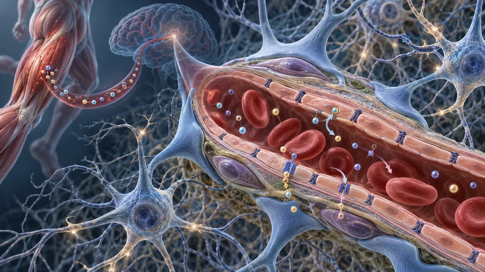
When exercise is described as protective for the aging brain, the discussion often turns quickly to neurons. A 2024 narrative review asks readers to look at the infrastructure around those cells as well: the blood vessels that deliver oxygen and nutrients, adjust circulation to neural activity and help keep the brain’s internal environment separate from the rest of the body.
The review, published July 1, 2024, connects findings from geroscience, animal research, observational studies and exercise interventions. Its contribution is not a new clinical trial showing that exercise preserves memory. It is an attempt to connect several levels of biology, from cellular aging to cerebrovascular function, and to show where the evidence is stronger, more indirect or still incomplete.
Why brain aging involves blood vessels
The brain depends on vessels that can respond when nearby neural tissue becomes more active. This requires the vessel lining, called the endothelium, to release signals that relax the vessel and adjust local blood flow. The review also emphasizes the blood–brain barrier, a selective interface that helps regulate what moves from the circulation into brain tissue.
Aging can disrupt both functions. The review describes how cellular aging and cardiovascular risk factors such as obesity, diabetes and hypertension can contribute to vascular dysfunction. When endothelial cells become senescent, they remain present but no longer behave like healthy, responsive cells. They produce less nitric oxide, a molecule involved in vessel relaxation, which can weaken the blood-flow response to neural activity.
Senescence can also affect the blood–brain barrier. The review links the accumulation of senescent vascular cells with impaired barrier integrity, loss of supporting pericyte and transporter functions, and less effective removal of waste products. A compromised barrier may allow inflammatory signals from the circulation to enter an environment that normally has tighter protection. The possible consequences include neuroinflammation and impaired neuronal or glial function.
This is the vascular connection in practical terms. Brain aging is not only about what happens inside brain cells. It may also involve whether the vessels can respond to changing energy demands and maintain the boundary that supports the brain’s chemical stability.
Exercise can change the signals vessels receive
Exercise may influence this system through more than one timescale. During an individual bout, changes in cardiac output and cerebral blood flow can alter the forces and signals experienced by vessels. Repeated training may also produce longer-lasting adaptations in cellular energy handling, inflammation and endothelial function. The review treats these immediate physiological effects and slower biological changes as related but not interchangeable.
One example involves AMPK, an energy-sensing pathway activated by moderate-to-heavy exercise. In the review’s account, AMPK helps conserve cellular energy and activates PGC-1α, which supports mitochondrial biogenesis. Mitochondria are the structures that help cells produce energy, while mitophagy removes damaged mitochondria. Aging-related declines in these processes can increase oxidative stress, inflammation and endothelial dysfunction.
The proposed vascular relevance is broader than energy production alone. AMPK can reduce inflammatory transcriptional activity in blood vessels, support nitric-oxide-dependent relaxation and promote angiogenesis through vascular endothelial growth factor, or VEGF. These mechanisms could help vessels respond to the brain’s metabolic demands, but the review presents them as connected biological possibilities rather than a fully demonstrated human sequence from exercise to preserved cognition.
Muscle-to-immune communication offers a complementary example. Exercise causes muscle to release interleukin-6, or IL-6, in a context different from the IL-6 released by immune cells such as macrophages. The review describes muscle-derived IL-6 as a myokine, a signal produced by muscle that can stimulate anti-inflammatory interleukins including IL-10 and IL-1 receptor antagonist. Those signals can suppress the pro-inflammatory cytokine TNF-α.
Over time, this type of communication may help protect vascular and blood–brain barrier function. It illustrates why exercise research cannot be reduced to a single “brain chemical.” The proposed effects involve interactions among muscle, blood vessels, immune signaling and cellular energy systems. The important question is how consistently those interactions appear in humans and whether their vascular consequences last long enough to support thinking and memory.
What human studies actually measured
The human evidence summarized by the review covers several different outcomes, and they should not be treated as interchangeable.
One large meta-analysis included 51 randomized controlled trials involving 2,260 middle-aged and older adults and reported improvements in endothelial function after exercise. This is useful evidence that exercise can affect how the vessel lining behaves. It is not the same as 51 trials directly demonstrating improved brain circulation, slower cerebrovascular aging or better cognition. Endothelial function is a vascular measurement that helps illuminate a mechanism; it is not itself a memory outcome.
A different kind of evidence comes from observational comparisons. Lifelong exercisers who maintained higher cardiorespiratory fitness into older age had higher cerebral blood flow and cerebrovascular reactivity than sedentary counterparts in the studies described by the review. Cerebrovascular reactivity refers to the ability of brain vessels to adjust their response under a physiological challenge. These findings connect long-term fitness with measurements of brain circulation, but comparisons between people who exercise throughout life and those who do not can reflect many differences beyond exercise itself.
Shorter training studies ask another question. The review cites evidence that 12 weeks of exercise can improve cerebral hemodynamics and cerebral blood volume in the dentate gyrus of healthy older adults. Cerebral hemodynamics describe the movement and regulation of blood through the brain, while cerebral blood volume describes how much blood is present in a region at a given time. In related animal work, increases in dentate-gyrus blood volume were correlated with neurogenesis.
These studies are valuable because they suggest that brain circulation may remain responsive to training, including later in life. They still measure intermediate outcomes. A change in blood flow, vascular reactivity or regional blood volume can help explain how exercise might support brain function without becoming evidence that dementia has been prevented or that memory will remain preserved for a particular person.
VEGF shows why location matters
VEGF provides a useful test of how carefully these findings need to be interpreted. It is a growth-related signal involved in the formation and maintenance of blood vessels. Exercise studies in humans have reported increases in VEGF in serum or plasma, while other studies in older adults found no effect. The review links these mixed results to differences in exercise modality, intensity and participant characteristics.
The biological compartment matters too. Systemic VEGF in blood does not automatically tell us what happens inside the brain. The review notes that whether VEGF in the circulation can cross the blood–brain barrier remains unresolved. Animal studies have reported exercise-related increases in hippocampal VEGF, while human evidence from the brain is more limited.
One human finding came from older patients with normal-pressure hydrocephalus. After an acute session of moderate-intensity upper-body resistance exercise, cerebrospinal fluid VEGF increased in the exercise group compared with controls. Cerebrospinal fluid is closer to the brain than blood plasma, so the result raises a different biological question from a blood measurement. It still does not establish that new brain vessels formed, that the response occurs in other older adults, or that it preserves cognition.
VEGF therefore helps clarify rather than simplify the story. The same named molecule can be measured in different places, after different kinds of exercise and in different populations. A result in one compartment or participant group cannot automatically be transferred to another. Nor can a growth-factor response, by itself, settle the larger question of lasting cognitive benefit.
Lifelong fitness is not the same as starting later
One of the review’s most important boundaries concerns timing. People who maintain fitness across adulthood may have a different vascular history from people who begin exercising after age-related cellular and vascular changes are already present. Observations about lifelong exercisers cannot answer whether starting later produces comparable protection.
The available research also has design limits. Much of the literature discussed in the review is cross-sectional or observational, and a substantial portion comes from animal models. These approaches can reveal associations and suggest mechanisms, but they cannot establish the full causal chain in humans. Positive findings may also be more likely to appear in the published literature than null or negative results. In addition, many populations and combinations of age, ethnicity and disease are not adequately represented.
The review’s authors therefore leave open several practical scientific questions. Researchers still need to understand how exercise frequency, intensity, duration and modality relate to cerebrovascular and cognitive outcomes in older adults. They also need to determine whether the vascular changes associated with lifelong fitness can be reproduced after later-life training begins. Those are questions about timing and applicability, not invitations to infer an optimal exercise prescription from this review.
What healthier brain vessels could mean for aging
For older adults, the possible benefit is a brain whose vessels remain better able to adjust blood flow and maintain the blood–brain barrier as demands change. Exercise strategies that preserve those functions could help sustain thinking and memory with age, but only if the relevant vascular adaptations translate into lasting cognitive benefit in humans and can be produced after the point when many people begin exercising.
That conditional path is the review’s real significance. It shifts attention toward the blood-vessel system supporting the brain while keeping the distance between a plausible mechanism, a measured vascular change and a meaningful cognitive outcome visible. Establishing when exercise can create durable vascular responsiveness, and for whom, remains necessary before the proposed protection can become a reliable promise for aging brains.
Key Sources
- Primary source: Exercise to Mitigate Cerebrovascular Aging: A Geroscience Perspective.
DOI or Journal Page · Review or Meta-Analysis · 2024-07-01
Keep exploring