
Research Signal – Source date 2025-06-26
A laboratory model can show that a drug affects colorectal cancer cells. But how much does that result say about whether the same treatment will help a person? The answer depends on what the model includes—and leaves out.
A review published June 26, 2025, compares cell cultures, animal models, and newer platforms for studying colorectal cancer. Its literature search covered English-language research indexed through January 2025 in PubMed/MEDLINE, Scopus, and Web of Science. The review appraises how these systems can support different research questions; it is not a human trial testing model-guided treatment.
A cell line can isolate a useful drug question
Flat, two-dimensional cell cultures are popular in part because they are accessible and allow researchers to examine cancer cells under controlled conditions. A panel of cell lines can also represent different tumor mutations, helping researchers ask whether a drug’s effect changes with a cancer’s molecular features.
For example, the review describes Caco-2 cells as KRAS- and BRAF-wild-type, while several other commonly used lines carry KRAS mutations and HT29 carries a BRAF mutation. These differences matter when investigating drugs that target the epidermal growth factor receptor, or EGFR. The review explains that EGFR-targeted drugs such as cetuximab and panitumumab are used in contexts where tumors lack RAS or BRAF mutations. In cell-line experiments, Caco-2 can serve as a model predicted to be sensitive to EGFR inhibition, while lines with the relevant mutations can help researchers study resistance.
This is a focused use of a model: select cells with defined molecular features, then test a mechanism or drug response against that background. The result can help clarify why a treatment may behave differently across cancers. A flat culture, however, gives every cell similar access to nutrients and drug in the surrounding medium. It also lacks the gland-like architecture found in colorectal tumors, limiting what it can reveal about how tissue structure shapes a response.
A sphere changes the conditions around the cells
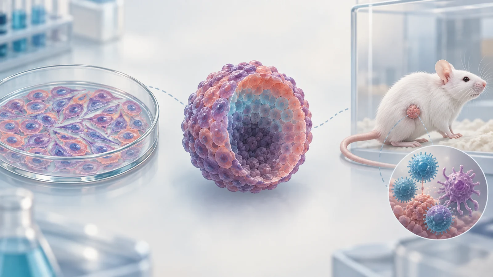
When cancer cells grow as a three-dimensional spheroid, they form a compact structure with cell-to-cell contacts. As the sphere grows, oxygen and nutrients may become less available toward its center, and drug access can vary across its layers. Some spheroids develop an outer proliferating zone and a more quiescent inner core; larger ones may develop a necrotic center.
Those gradients change the experiment. In a flat dish, a drug reaches cells across the surface in broadly similar conditions. In a spheroid, researchers can investigate how a treatment performs when cells occupy different physical and metabolic environments. The review describes differences in growth, gene expression, and drug response between three-dimensional cultures and monolayers, making spheroids a bridge between simple cell-line tests and more complex systems.
A spheroid still represents a limited slice of a tumor. Its value lies in adding architecture and uneven access to the question, not in reproducing every feature of a person’s cancer.
Mouse models trade human tumor cells for different kinds of context
A human tumor graft in an immunodeficient mouse preserves human cancer material, but the host’s immune system is deliberately limited so it does not reject the graft. This can support studies using patient-derived tumors, while making the setting less suited to examining a complete immune response to those tumors.
A syngeneic model makes a different trade. Mouse colorectal cancer cells, such as CT26 or MC38, are transplanted into a mouse of the matching genetic background. The immune system remains intact, giving researchers a way to study tumor–immune interactions and treatments such as checkpoint inhibitors. The cancer cells and host are both murine, though, rather than a patient’s tumor and human immune system.
Humanized mice aim to bring some human immune components into an immunodeficient mouse. Researchers can reconstitute these mice with human blood-forming stem cells or peripheral blood immune cells and then study a human tumor graft in that setting. The arrangement permits investigation of interactions between human immune cells and colorectal cancer, while remaining a constructed model rather than a person’s own immune environment.
Each design changes which part of the biology is easiest to observe. The choice depends on whether the central question concerns human tumor material, an intact immune system, or interactions involving human immune cells.
Engineered tumors and induced disease answer different questions
Genetically engineered mouse models can start with defined cancer-related mutations and follow tumor development in an animal. The review describes the ApcMin/+ mouse, in which an Apc mutation drives intestinal adenomas. But these growths occur mostly in the small intestine and rarely progress to invasive cancer, unlike the main disease features researchers may want to study in human colorectal cancer. More complex combinations of engineered changes can produce tumors that progress further, though the resulting models still represent selected genetic routes to disease.
Chemically induced models pose another kind of question. The review discusses tumors produced with azoxymethane, sometimes alongside dextran sodium sulfate, which causes intestinal inflammation. These systems can be useful for studying tumor formation in an inflammatory setting. Their mutation patterns and disease course can differ from human colorectal cancer, and the inflammation itself can affect the outcome. The review also describes limited metastatic behavior in these models, constraining their use for questions about spread to distant organs.
So a model that captures a defined driver or inflammatory process may be useful for studying that process, even if it does not recreate the location, progression, or spread of a person’s tumor. Model selection shapes the question that an experiment can answer.
Researchers are testing ways to connect the platforms
The review presents organoids, tumor-on-chip systems, humanized mice, and computational methods as parts of a broader effort to bring more patient-relevant features into experiments. A chip can incorporate flow and other physical conditions, while computational approaches can combine measurements such as molecular and imaging data. These platforms offer ways to pose questions that a static cell culture cannot address alone.
The authors propose an iterative research path: use cell-based experiments to investigate a mechanism, test findings in a chip or other system with additional context, and then evaluate selected results in an animal model. Computational tools could help prioritize which treatments or mechanisms to examine. The purpose is to make the next experiment more informative by asking whether an idea holds across different biological settings.
Examples discussed in the review illustrate the appeal of that approach, including work combining patient-derived material, chips, and mouse models. They do not amount to a common prospective test showing that a combined workflow can reliably forecast an individual patient’s treatment response. Nor do discussions of AI, chips, or “avatars” establish routine clinical use or measured benefit for patients. Those possibilities depend on whether predictions made across platforms prove reliable against human outcomes.
What better tumor models could mean for treatment choices
If a combined set of models can reliably predict treatment response in people with colorectal cancer, clinicians and patients might one day use those predictions to choose among available treatments. That use remains hypothetical: prospective studies would need to show that predictions made before treatment match patients’ outcomes and that acting on them improves care.
Key Sources
- Primary source: Exploring Experimental Models of Colorectal Cancer: A Critical Appraisal from 2D Cell Systems to Organoids, Humanized Mouse Avatars, Organ-on-Chip, CRISPR Engineering, and AI-Driven Platforms-Challenges and Opportunities for Translational Precision Oncology.
DOI or Journal Page · Review or Meta-Analysis · 2025-06-26
Keep exploring