
Research Signal – Source date 2024-08-01
Solid tumors create a difficult testing problem for engineered immune-cell therapies. CAR T cells have produced striking results in some blood cancers, but solid tumors can be harder to reach and may contain conditions that limit T-cell activity. Researchers also need to decide which tumor target and CAR design deserve more extensive testing. That work often relies on animal models that take substantial time and resources to establish, making it difficult to compare many candidates quickly.
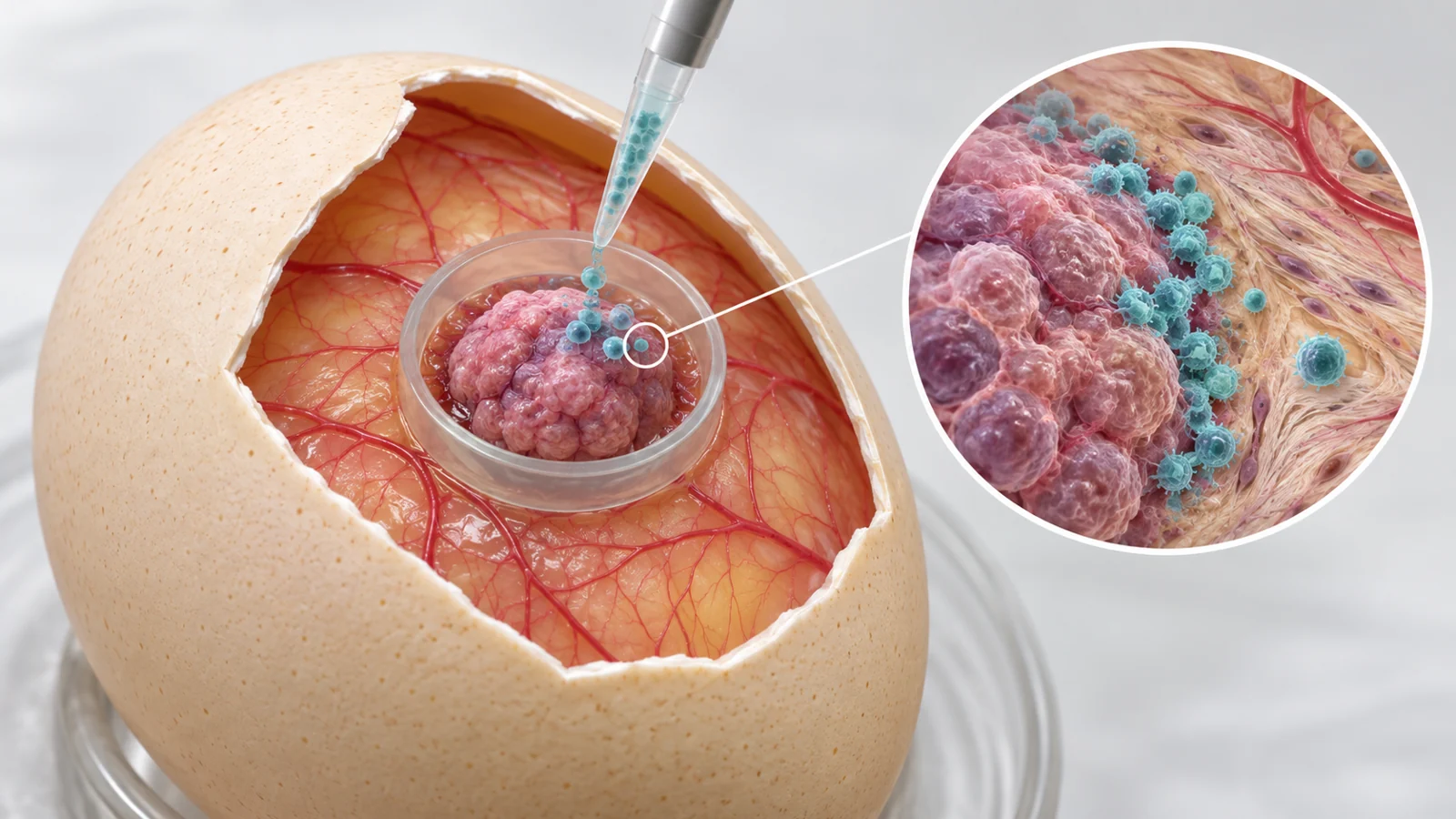
A study published on August 1, 2024, tested whether a fertilized chicken egg could provide a useful intermediate setting. The experiment used the chorioallantoic membrane, or CAM, a highly vascularized membrane beneath the shell that supports the developing embryo. Human tumor cells placed on this membrane formed three-dimensional grafts that could be treated and examined over roughly a week. The result is not a human treatment trial. It is a preclinical experiment asking what targeted T cells do in living, vascularized tumor tissue over a short observation period.
A living membrane turns an egg into a short-term tumor model
The CAM naturally transports gases, nutrients, and calcium to the chick. Its blood-vessel-rich tissue can also support engrafted cells and tumor tissue. In this study, three human cancer cell lines were placed on the membrane. FaDu and SCC-47 were head-and-neck squamous cell carcinoma lines that expressed HER2, the cell-surface protein selected as the CAR T-cell target. MDA-MB-468 was a breast adenocarcinoma line that did not express HER2.
The researchers confirmed that all three lines established tumors on the CAM. They used tissue staining to identify the grafts and to examine the surrounding membrane, where vascularization was evident. The tumors also carried a luminescent reporter, allowing the investigators to follow changes in the signal over time as an estimate of viable tumor burden.
That combination matters because the CAM adds features missing from simpler cell cultures. Spheroids and organoids can form three-dimensional structures, but they are not vascularized in the same way as a graft growing on a living host membrane. At the other end of the preclinical spectrum, mouse xenografts can provide a more demanding in vivo setting but generally require more time and care. The CAM was being tested as a practical middle step, not as a complete substitute for either approach.
The experiment tested target matching over four days
The treatment sequence was direct and limited. After tumor cells were engrafted, the researchers waited three days and then applied the treatment suspension directly onto each growing tumor within its ring on the CAM. One group received HER2-specific CAR T cells. The control group received expanded T cells that had not been given the CAR. Four days after treatment, the eggs were examined again, so the reported treatment window ran from day three to day seven after engraftment.
The CAR T cells were engineered to recognize HER2. That made the HER2-negative MDA-MB-468 graft an important comparison: it helped test whether the treatment-associated changes were linked to the chosen target rather than to exposure to T cells alone. The researchers used luminescence imaging before treatment and at the later assessment, then compared the growth patterns between treatment groups. They also examined tissue for pan-cytokeratin and Ki-67. Pan-cytokeratin helped identify tumor tissue, while Ki-67 provided a measure related to cell proliferation.
The design therefore asked a focused question. When the transferred T cells encounter a tumor carrying their selected surface target, does the short-term tumor signal and tissue appearance change more than with control T cells or with a tumor lacking that target?
Targeted treatment changed growth, but not uniformly
The growth-pattern slopes differed between CAR T-cell-treated and control-treated tumors for both HER2-positive lines, FaDu and SCC-47. The corresponding difference was not observed in the HER2-negative MDA-MB-468 tumors. That pattern supports target-dependent activity within the boundaries of this experiment: the engineered cells were associated with different growth behavior when the tested target was present.
The size of the response also varied between the two HER2-positive lines. FaDu showed the particularly pronounced reduction in luminescence from viable tumor cells by the later measurement. SCC-47 showed a treatment-associated difference as well, but the effect was less dramatic in that readout. Tissue staining added another layer of evidence: both HER2-positive tumor types had lower Ki-67 staining after CAR T-cell treatment than their control-T-cell counterparts.
Similar HER2 expression did not produce identical measured responses. The authors suggest that differences between the tumor lines, including their biological characteristics and growth behavior, could help explain why the same targeting strategy had a stronger visible effect in FaDu. A target can make a tumor recognizable without making every target-positive tumor equally vulnerable over the same short interval.
The T cells gathered where tumor met supporting tissue
The investigators also asked where the transferred cells were located, not only whether the tumor signal changed. In a separate localization experiment, they used a higher effector-to-tumor ratio to make the T cells easier to identify in tissue sections. CD3 staining detected T cells four days after treatment, supporting their persistence in the CAM and tumor tissue during the observation period.
The cells were not distributed evenly throughout the grafts. In both HER2-positive FaDu tumors and HER2-negative MDA-MB-468 tumors, most of the detected T cells accumulated at the interface between epithelial tumor nests and the surrounding CAM stroma. The same location in the target-negative model is important: physical access to the graft and persistence in its tissue are not identical to target-dependent killing.
This spatial result raises a practical question about how engineered cells reach tumor cells inside a solid mass. It is consistent with an access problem at the boundary of tumor and supporting tissue, but the staining does not establish that the CAM reproduces human immune exclusion or that every detected T cell was functionally active. The treatment was also applied directly to the tumor. The experiment therefore informs local exposure and tissue location, not how these cells would traffic after intravenous administration.
A useful middle ground with important missing pieces
The CAM can support questions that a dish-based assay cannot. Researchers can compare tumor burden over several days, inspect tumor architecture, observe whether transferred T cells remain in the tissue, and test how a locally applied CAR T-cell candidate behaves in a vascularized graft. Those observations may help narrow the field of designs that merit deeper study.
The model also leaves important biology outside the experiment. The four-day post-treatment assessment cannot establish durable tumor control, recurrence prevention, or long-term T-cell persistence. Human donor cells and a previously studied CAR construct do not turn the experiment into evidence of clinical efficacy or safety. Nor does this study establish agreement with patient outcomes, quantified throughput or cost advantages, or replacement of mouse-based validation.
The authors describe several ways the system could be expanded. CAM tumors could be supplemented with cells associated with an immunosuppressive tumor environment, including regulatory and stromal populations, to test combination treatments and additional immunotherapies. Patient-derived tumor grafts could also be explored for individualized treatment sensitivity testing. Those are proposed extensions, not outcomes demonstrated by the three cell-line grafts in this paper.
What faster testing could mean for solid-tumor therapies
At the time of publication, the practical promise of the CAM approach was to give researchers another living setting in which to examine engineered immune-cell designs before committing them to more demanding preclinical studies. A dependable intermediate test could make it more practical to investigate a wider range of CAR T-cell candidates in vascularized tumor tissue and carry the most informative candidates into deeper development.
That consequence remains conditional. The model would need reproducible performance across additional tumor types, CAR designs, and experimental settings, along with evidence that its short-term responses help predict which candidates deserve subsequent testing. If those conditions are met, the approach could help develop CAR T-cell treatments better matched to particular solid tumors, with the aim of achieving more effective tumor control. Any such patient benefit would still require further preclinical validation and clinical trials establishing efficacy and safety.
Key Sources
- Primary source: Chick Embryo Chorioallantoic Membrane as a Platform for Assessing the In Vivo Efficacy of Chimeric Antigen Receptor T-cell Therapy in Solid Tumors.
DOI or Journal Page · Peer-Reviewed Paper · 2024-08-01
Keep exploring