
Research Signal – Source date 2026-06-04
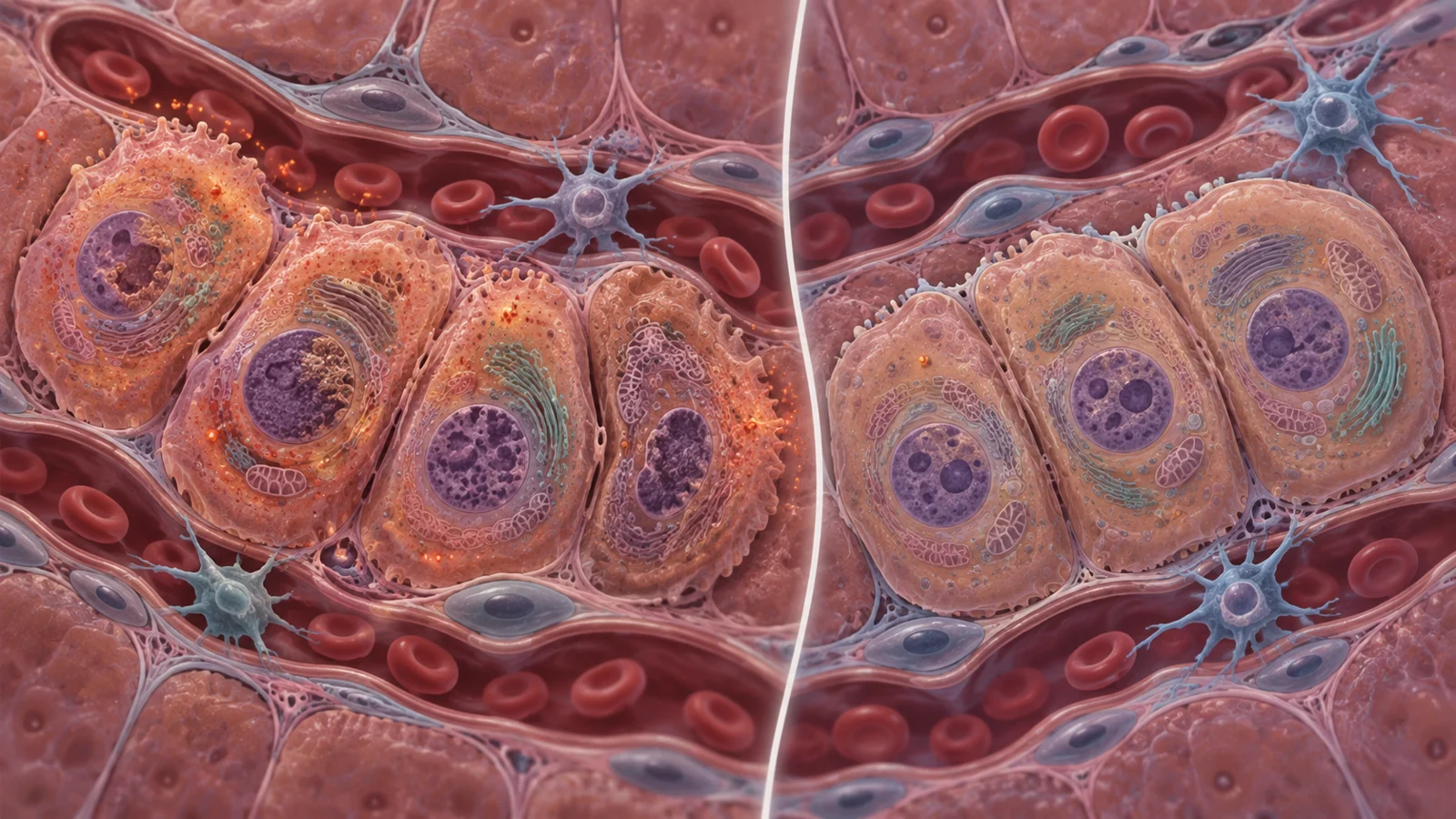
A mouse experiment found that prolonged cell resetting was followed by severe liver injury and early death. The liver showed signs of oxidative stress, a damaging imbalance involving reactive oxygen species. Giving the mice the antioxidant N-acetylcysteine delayed death, while measured markers of the cell-state change remained. The result points to oxidative stress as part of the danger, but it does not show that the treatment prevented liver failure or preserved useful regeneration.
The study used four factors—OCT4, SOX2, KLF4 and c-MYC—to push mature cells toward a more flexible state. This process, called cellular reprogramming, resets aspects of a cell’s identity. The factors are often abbreviated as OSKM. Here, researchers induced them throughout mice using doxycycline in their drinking water, then examined how sustained exposure affected the liver.
Liver injury appeared within days
After three days of continuous factor induction, liver cells had lost markers associated with mature hepatocytes, the liver’s main working cells. At the same time, the researchers found increased Sox9, a marker associated with liver progenitor-like cells. The pattern was consistent with hepatocytes shifting toward a less mature state. Markers of DNA damage and apoptosis, or programmed cell death, also rose.
The animals’ condition worsened quickly. Four days of continuous induction brought rapid weight loss and premature death. Blood tests showed elevated AST and ALT, enzymes that rise when liver cells are injured. These findings link the short exposure to acute liver damage; they do not establish that every cell changed identity in the same way or that the shift produced functioning replacement tissue.
The timing also mattered. When doxycycline was withdrawn, the mature-cell marker HNF4α returned within 24 hours, alongside a marked reduction in cells positive for OCT4. That quick reversal suggests the observed change in liver-cell state was not necessarily permanent. Meanwhile, continued induction was associated with accumulating injury.
Stress signals came from the liver cells
Reactive oxygen species, or ROS, are chemically reactive molecules produced in cells. In excess, they can damage cellular components. The researchers found more ROS-positive hepatocytes after OSKM induction, along with increased DNA-damage markers, apoptosis and stress signaling. Isolated liver cells also showed increased NRF2 protein and activity in genes that respond to oxidative stress.
NRF2 is part of a cellular defense response that helps regulate antioxidant activity. Its activation, by itself, would show that cells sensed stress, not that oxidative stress caused the animals’ deaths. But several observations converged: ROS increased in the liver cells, stress-response genes rose over time, and liver injury appeared alongside those signals. Analysis of previously generated single-nucleus RNA sequencing data also found oxidative-stress and NRF2-pathway activity in liver progenitor-like cells during OSKM induction.
The comparison with acetaminophen-related liver injury added context. Both settings included liver progenitor-like cells, but the OSKM-associated cells showed stronger enrichment of ROS and oxidative-stress pathways. The researchers therefore proposed that sustained reprogramming created a redox burden that contributed to liver damage. These data support that explanation, while leaving room for other processes to contribute to the injury.
Similar factor levels, different stress burden
Female mice were more resistant than age-matched males to premature death during continuous induction. The difference did not track with a clear difference in the measured OSKM expression or liver-cell dedifferentiation: both were similar between the sexes. Instead, female mice had fewer ROS-positive hepatocytes, and their liver cells had higher baseline expression of some antioxidant and glutathione-related genes.
The sex comparison helps separate the amount of cell-state change from the amount of oxidative stress observed. Some NRF2-responsive genes differed between males and females, while others were induced in both. The results suggest that differences in antioxidant defenses may influence how well liver cells tolerate the same reprogramming pressure. They do not establish a sex-specific response in people or explain every factor behind the mice’s survival difference.
An antioxidant delayed death while markers remained
The researchers tested N-acetylcysteine, or NAC, as a way to reduce oxidative stress. With NAC, premature death was significantly delayed. The treatment also reduced liver-cell DNA-damage markers and NRF2 nuclear accumulation. Meanwhile, measured OSKM expression and reprogramming-associated markers, including Sox9, largely remained.
This intervention strengthens the case that oxidative stress contributed to the harm: lowering stress signals coincided with improved survival. Yet the rescue was partial, described as delayed lethality rather than prevention of death. And retaining cell-state markers is not the same as demonstrating that the liver regenerated, worked better or reversed aging. NAC served as an experimental probe in mice; these findings do not establish a human treatment or dose.
Exposure time may shape the tradeoff
The experiment raises a practical question for future reprogramming research: can exposure be brief enough to encourage cell plasticity while giving tissues time to recover? In this study, withdrawal was followed by a rapid return of a mature liver-cell marker, while sustained induction was accompanied by worsening injury. That makes duration a plausible factor to investigate.
The authors point to cyclic induction as a possible strategy, but this experiment did not directly compare a cyclic schedule with continuous exposure. Nor did it establish that a particular schedule would preserve durable repair while avoiding harm. The central challenge is to distinguish a temporary marker shift from a lasting, beneficial change in tissue function.
What liver protection could make possible
If future work can limit liver injury while preserving meaningful regenerative effects, controlled cell reprogramming might become more usable as a way to support tissue repair. The path depends on finding an effective exposure schedule that changes cell state without overwhelming the liver’s defenses. This mouse study’s survival delay and marker results alone do not deliver that capability; they identify oxidative stress as a problem that future approaches would need to solve.
Key Sources
- Primary source: Oxidative stress drives liver failure during in vivo partial reprogramming.
DOI or Journal Page · Peer-Reviewed Paper · 2026-06-04
Keep exploring