
Research Signal – Source date 2026-04-21
Cardiovascular disease remains the world’s leading cause of illness, disability and death. The review published on April 21, 2026, notes that the global number of people living with cardiovascular disease rose from 271 million in 1990 to 523 million in 2019, while related deaths increased from 12.1 million to 18.6 million. Treatments have become more effective, but risk can remain even after conventional risk factors and diagnosed conditions are addressed.
The review asks whether some of that remaining risk could be approached by targeting cardiovascular aging itself. Its contribution is an organizing framework: it brings molecular damage, cell behavior, organ-level changes and wider physiological influences into one account. It does not show that aging has been reversed in patients. Instead, it clarifies why preserving the aging heart may require more than treating one disease at a time.
Aging is more than elapsed time
Cardiovascular aging is not a single clock running at the same speed in every person or tissue. The review groups its twelve hallmarks across molecular, cellular and systemic levels. Its discussion connects mitochondrial dysfunction and oxidative stress with inflammation and cellular senescence. At the systemic level, it includes the renin–angiotensin–aldosterone system, β-adrenergic signaling, growth signaling and mechanosignaling.
These levels interact. A change in vessel mechanics can alter signaling; damaged mitochondria can intensify inflammation; inflammatory signals can affect neighboring cells and the wider circulation. The value of the framework is therefore less a catalogue of aging features than a way to connect processes that are often studied separately.
The clinical motivation is practical. A person may receive treatment for hypertension, diabetes, dyslipidemia or another cardiovascular condition and still face declining vascular flexibility, impaired perfusion or reduced cardiac reserve. Aging biology could become an additional therapeutic target, but the same interconnectedness that makes it attractive also makes broad intervention difficult to interpret.
The blood supply changes at two scales
Large arteries and the small vessels that feed heart muscle do not age in exactly the same way.
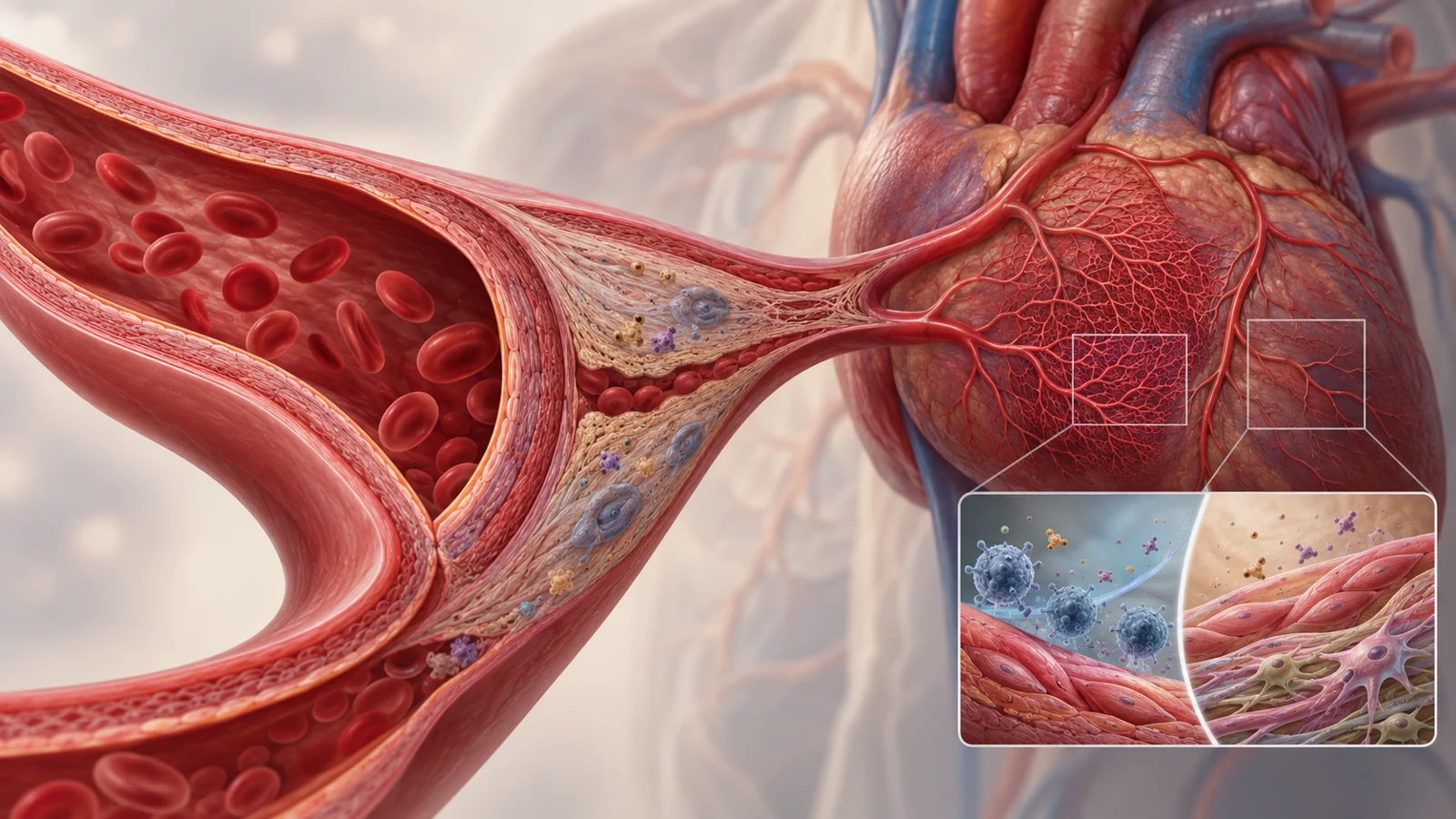
In larger vessels, aging is associated with remodeling of the vessel wall. The review describes dilation of the lumen, thickening of the intima and media, chronic inflammation, calcification, arterial stiffening and reduced elasticity. A stiffer artery changes how pressure and blood flow are handled across the circulation. Vascular cells may also become dysfunctional or senescent, weakening the normal balance that keeps the vessel responsive.
The heart’s microvasculature presents a different problem. The coronary microcirculation can lose capillary density while its vessel walls become stiffer and thicker. Endothelial dysfunction reduces the availability of nitric oxide, a molecule involved in vessel relaxation. Coronary blood flow and coronary flow reserve can also decline. Coronary reserve describes the capacity to increase blood delivery when the heart needs more oxygen; a lower reserve leaves less room to respond to demand.
These changes matter because the heart depends on a dense, adaptable network of small vessels, not only on open major arteries. Reduced capillary density and impaired small-vessel relaxation can limit oxygen and nutrient delivery to cardiomyocytes. Increased vascular permeability and lymphatic dysfunction may add to the difficulty of maintaining a stable tissue environment. The result is a physical loss of flexibility in the route from blood pressure to heart-muscle supply, even when the relevant problem is too small to be described simply as a blocked large artery.
One chain links oxidative stress with inflammatory spread
The review describes a concrete biological chain. During vascular aging, NADPH oxidase can produce more reactive oxygen species. Excess reactive oxygen species contribute to inflammatory signaling and degrade nitric oxide or reduce its effective availability. With less nitric oxide, the endothelium becomes less able to promote vessel relaxation, encouraging vasoconstriction and poorer tissue perfusion.
Oxidative stress and inflammation can then reinforce one another. The review highlights activation of the NF-κB pathway and describes how reduced endothelial nitric oxide synthase activity contributes to endothelial dysfunction. In the heart, lower nitric oxide signaling is also linked in the review to impaired contractile and diastolic function through a PKG-dependent pathway.
Cellular senescence adds another route for local damage to spread. Senescent cells release a mixture of cytokines, chemokines, growth factors and matrix metalloproteinases known as the senescence-associated secretory phenotype, or SASP. These secretions can influence nearby cells and induce secondary senescence. The cGAS–STING pathway is one connection between cytosolic DNA sensing, inflammatory signaling and the development of senescence.
This local biology sits inside the review’s wider systemic picture. Chronic changes in hormonal, neural, growth and mechanical signaling can keep cardiovascular tissues in an altered state. Targeting one node may therefore change several downstream processes, but it may also affect functions that the body still needs for repair and adaptation.
Cardiometabolic health, environment and behavior leave different traces
The review treats cardiovascular aging as the product of several overlapping influences. Cardiometabolic disorders are one example. In a cross-sectional analysis of 39,559 UK Biobank participants, machine-learning methods were used to estimate cardiovascular age, and hypertension, diabetes and dyslipidemia were collectively associated with faster cardiovascular-aging trajectories. This kind of finding can reveal a population pattern, but it does not determine an individual’s aging rate or by itself prove that one condition caused the measured trajectory.
Environmental exposure provides another layer. The review cites analysis of 3,772 National Health and Nutrition Examination Survey participants in which chronic exposure to metals including cadmium, cesium, cobalt and lead showed dose-dependent associations with vascular-aging biomarkers. It also discusses experimental evidence in which chronic low-dose radiation altered cardiac protein patterns related to energy metabolism, extracellular-matrix regulation, oxidative stress and senescence-associated signaling. Human observations and tissue experiments answer different questions, so they should not be treated as interchangeable evidence.
Behavior and diet can influence the same broad biology through still different kinds of evidence. Higher diet-quality scores and adherence to Mediterranean dietary patterns are associated with more favorable vascular-aging profiles in the review. Physical activity, less sedentary behavior and weight management are also described as ways to improve vascular-aging phenotypes. These observations fit with mechanistic concerns about metabolism, inflammation and vessel function, but they do not turn the review into a personal prevention plan or establish one universal intervention for every older adult.
Why the same regulator can have different effects
Aging-associated regulators illustrate why treating a process is not the same as simply suppressing it.
In mouse models of pressure overload, increased p53 signaling promoted cardiac dysfunction. The review links p53 to inhibition of hypoxia-inducible factor 1α and vascular endothelial growth factor, suppressing cardiac blood-vessel growth. It separately describes endothelial p53 experiments involving capillary loss, fibrosis and inflammatory-cell recruitment. Depleting endothelial p53 in those experimental settings improved capillary density and cardiac function. The findings make p53 an important biological regulator to investigate, but they do not establish that broadly reducing p53 would be safe or beneficial in people.
The p21 pathway shows a different complication. p21 is downstream of p53 and can contribute to senescence and apoptosis. Levels of p21 rise in older mouse cardiac tissue, yet systemic depletion of p21 reduced capillary density and impaired contractile function in the cited experiments. A molecule can therefore be associated with senescence while also participating in a function that supports tissue maintenance. Its effect depends on the cell type, organ, disease state and intervention used.
These regulator experiments should be distinguished from interventions that clear senescent cells. Senolytic approaches aim to remove selected senescent cells, whereas changing p53 or p21 signaling alters how cells respond to stress, growth arrest or damage. Those are not interchangeable strategies. The authors note that clearing senescent cells could carry risks such as impaired tissue repair. They also identify the need for more specific markers than p16 alone and for better understanding of how different senescent cardiac cell populations affect function.
Aging biology can therefore offer useful targets without making every aging-associated signal an enemy. The therapeutic question is not only whether a process contributes to deterioration, but also what that process is doing in a particular cell at a particular moment.
Clinical studies test targets before they establish benefits
The review connects its biological discussion to clinical investigation by listing studies aimed at different parts of cardiovascular aging. A completed study, NCT05872139, examined mitochondrial-derived oxidative stress and vascular endothelial dysfunction in older nonexercisers, comparing placebo with mitoquinone mesylate. A separate study, NCT05301192, was listed as recruiting to investigate cardiovascular effects of angiotensin-(1–7), with saline as the comparison.
These examples represent different targets: one focuses on mitochondrial oxidative stress, while the other concerns a component of cardiovascular signaling. Their presence in the review’s clinical-trial table shows that such questions have entered human research. A listed status, however, is not an efficacy result. The table entries alone do not provide an effect size, establish clinical benefit or determine safety. Nor does a list of FDA-approved drugs in the review mean that those drugs are approved specifically to reverse cardiovascular aging.
The distinction matters because the field is moving from biological plausibility toward testing, not skipping directly to a proven anti-aging treatment. Human studies will need to show that a target can improve meaningful cardiovascular function while preserving the repair, adaptation and tissue maintenance that aging-associated pathways may still support.
What this could change
For older adults, the practical payoff would be treatments that help preserve the heart’s small-vessel blood supply and functional reserve alongside established cardiovascular care. Better microvascular delivery could help the aging heart maintain oxygen access and respond to demand, potentially reducing residual disease risk rather than addressing only one named condition.
That capability remains conditional. It depends on demonstrating benefit in people, identifying which cells and pathways should be changed, and avoiding damage to tissue repair. If those bottlenecks are solved, cardiovascular aging could become a way to protect declining system-wide capacity—not a promise to make an old heart biologically young.
Key Sources
- Primary source: Cardiovascular ageing: hallmarks, signaling pathways, diseases and therapeutic targets.
DOI or Journal Page · Review or Meta-Analysis · 2026-04-21
Keep exploring