
Research Signal – Source date 2023-03-28
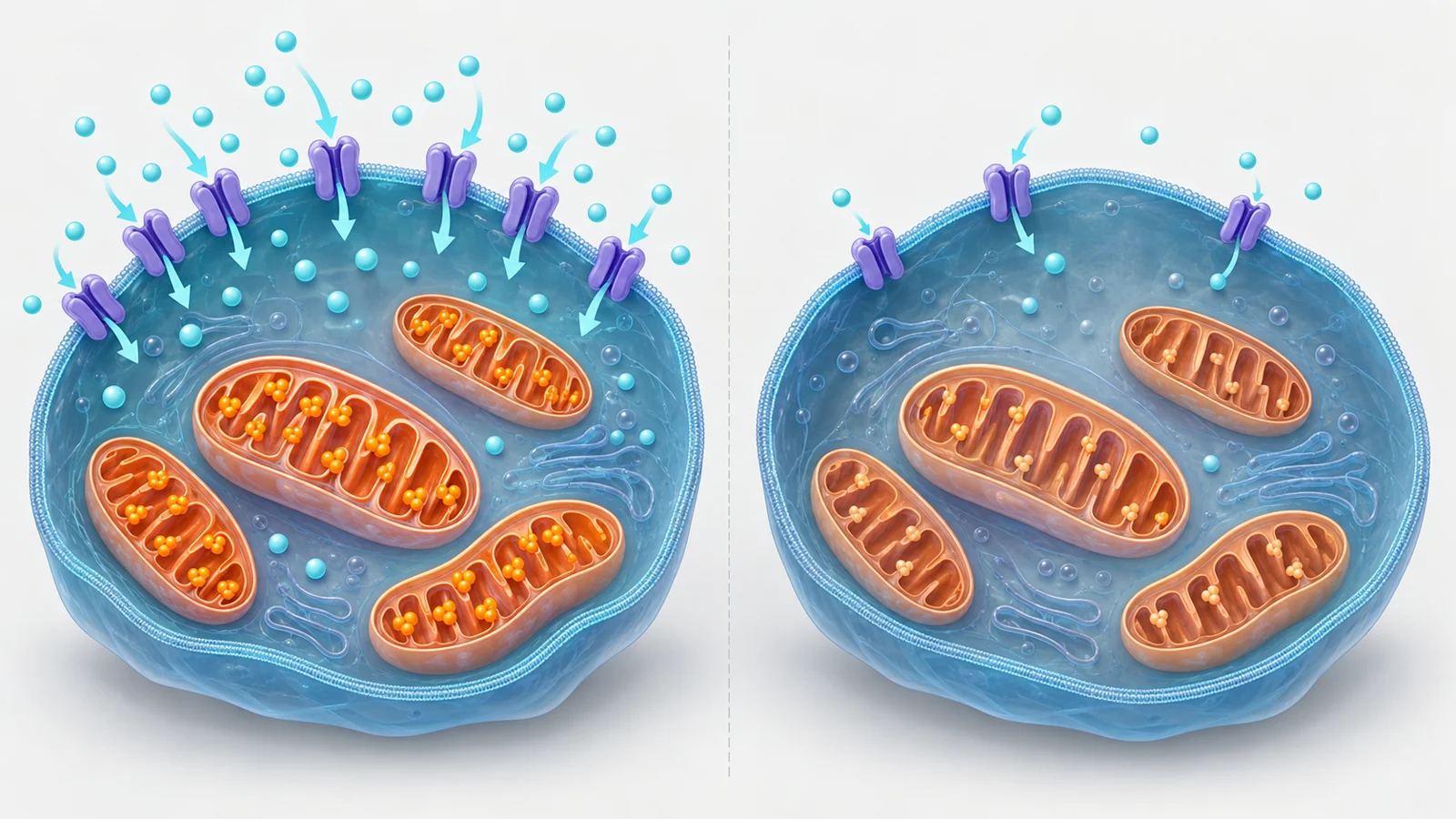
Creatine is part of the brain’s energy-buffering system, but getting it into cells depends on transport machinery. A March 28, 2023 study examined one possible influence on that machinery: cardiolipin, a fat concentrated in the inner membranes of mitochondria.
The experiment used growing hCMEC/D3 cells, a human-derived brain microvessel endothelial cell line. These cells are useful for studying properties of the blood-vessel lining that helps regulate exchange with the brain, but they are not an intact human blood-brain barrier. The researchers asked whether reducing cardiolipin would change the cells’ creatine transporter and their uptake of creatine.
A mitochondrial membrane fat meets a transport question
Cardiolipin helps organize mitochondrial membranes and supports proteins involved in energy production. It is not itself the creatine transporter. Creatine transporter-1, also called Crt1 and encoded by SLC6A8, is a membrane protein that helps move creatine into cells.
That separation matters. A change in a mitochondrial membrane lipid could affect a cell’s energy machinery, its membranes, or the expression of particular transport proteins. The study therefore measured several kinds of readout rather than treating creatine uptake alone as a complete explanation.
The publication also places creatine in the context of phosphocreatine, which can provide a rapidly available source for regenerating ATP during high energy demand. This makes transport into brain-related cells biologically interesting. It does not, by itself, show how much creatine reaches the living brain or whether changing transport would alter brain function.
What the researchers changed
The researchers used small interfering RNA, or siRNA, to suppress the cardiolipin-synthase-1 gene, hCLS1, for 48 hours. The enzyme catalyzes the final step in cardiolipin production. Mock-transfected cells served as the comparison group.
The intervention reduced hCLS1 levels by approximately 70% and cardiolipin levels by 35% relative to the mock-transfected controls. The team then followed several lines of evidence: messenger RNA for selected transporters, creatine-transporter protein, uptake of radiolabeled creatine, uptake of radiolabeled oleate, and cellular ATP production.
This sequence distinguishes the manipulation from the outcomes. The experiment did not simply add or remove creatine and observe a downstream response. It first altered cardiolipin synthesis, then tested whether transporter-related measurements and solute uptake changed in parallel.
The effect was selective rather than a general transport failure
Creatine-transporter mRNA fell by 45% after hCLS1 knockdown. The study also found lower creatine-transporter protein expression and reduced incorporation of radiolabeled creatine into the cells. The 45% figure describes the reduction in transporter mRNA, not the reduction in transporter protein or creatine uptake.
Other transporter transcripts did not all respond in the same way. Messenger RNA for P-glycoprotein and breast cancer resistance protein also decreased, while ABCC1, ABCC3, two MRP4 variants, and fatty acid transport protein-1 remained unchanged under the reported conditions.
Oleate uptake provided a functional comparison. It was unchanged after cardiolipin-synthase knockdown, consistent with the unchanged FATP1 messenger RNA. This pattern points to effects on selected transport systems rather than an indiscriminate loss of the cells’ ability to take up every tested solute.
The drug-transporter findings need their own boundary. Lower P-glycoprotein and breast cancer resistance protein mRNA identifies a change in gene expression, not a demonstrated change in drug transport, drug delivery, or treatment response.
Preserved total ATP narrows the interpretation
A simple explanation would be that the cardiolipin intervention weakened the cells so severely that they could no longer power membrane transport. The ATP measurements make that explanation incomplete under these assay conditions. Total cellular ATP production was unchanged, although the contribution from mitochondrial oxidative phosphorylation fell.
The result is informative because it separates total energy output from the balance of energy-producing pathways. The cells retained their overall measured ATP-production rate while showing a shift away from oxidative phosphorylation. That weakens the idea that reduced creatine uptake was merely a consequence of a whole-cell energy collapse.
It does not mean that every aspect of mitochondrial function stayed normal. Total ATP is an aggregate measurement, and the experiment does not resolve local energy availability at the transporter, short-lived metabolic changes, or every consequence of changing cardiolipin. The preserved oleate uptake also supports the authors’ view that the creatine result was not simply explained by generalized membrane damage.
What the experiment leaves unresolved
The observed chain is clear at the level of the measured cells: suppressing hCLS1 lowered cardiolipin, creatine-transporter mRNA and protein, and radiolabeled creatine uptake. The molecular connection between those steps is not clear. The authors explicitly leave open whether reduced cardiolipin affects membrane transport directly or through an indirect pathway.
The model adds another layer of caution. hCMEC/D3 is a transformed, high-passage cell line. It can reveal a response in a controlled setting, but growing cells in culture do not reproduce every feature of an intact blood-brain barrier, including the organization and interactions of living tissue.
Most importantly, uptake into these cells is not the same measurement as net creatine passage across the barrier. The experiment does not establish a rise or fall in creatine concentration in a living brain, and it does not test cognition, supplementation, or a clinical treatment. Nor does it show that increasing cardiolipin would reverse the transporter effect or be safe to attempt.
What this could mean for the brain’s creatine supply
If the relationship holds in the intact brain and its mechanism can be clarified, maintaining the cellular machinery that supports creatine uptake could become one route toward preserving access to a rapidly available energy buffer. The potential benefit would be better support for brain energy reserves, especially where endothelial transport contributes to the supply available to brain cells.
That possibility remains conditional on several unresolved steps. The cardiolipin–transporter relationship would need to operate in living brain-vessel tissue, the direct or indirect pathway would need to be understood, and any attempt to manipulate it would need to improve delivery safely. This cell experiment provides a reason to investigate that route; it does not provide a creatine dosing strategy, a cognitive benefit, or a treatment.
Key Sources
- Primary source: Reduction in cardiolipin reduces expression of creatine transporter-1 and creatine transport in growing hCMEC/D3 human brain microvessel endothelial cells.
DOI or Journal Page · Peer-Reviewed Paper · 2023-03-28
Keep exploring