
Research Signal – Source date 2022-07-01
Cancer treatment can succeed at its first job and still leave a lasting biological burden. As more children and adults survive cancer, clinicians see age-related diseases and loss of physical resilience appearing earlier than expected. The problems may include endocrine disorders, weakened bones, cardiac dysfunction, later cancers and frailty.
A review published on July 1, 2022, connects these clinical patterns with several possible mechanisms of biological aging. One candidate is cellular senescence, a state in which a cell stops dividing but remains metabolically active. The idea is not that senescence alone explains every late effect of treatment. It is that treatment-induced senescence may help connect an earlier exposure to damage that persists in normal tissues long after the cancer has been controlled.
Senescence keeps a damaged cell in place
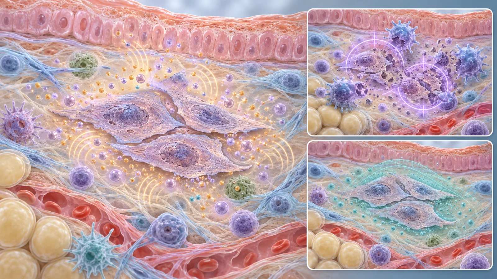
A senescent cell has entered growth arrest. Unlike a cell that has died, it remains present in the tissue. The review describes associated changes in chromatin, metabolism and protein production, along with increased resistance to programmed cell death. Senescent cells can therefore persist rather than being quickly removed.
Their effects may extend beyond the cell itself. Many senescent cells produce a senescence-associated secretory phenotype, commonly called the SASP: a mixture of inflammatory cytokines, chemokines, growth factors and enzymes. These signals can act on the senescent cell and on neighboring cells, changing the local tissue environment. A small population of persistent cells may consequently have effects that reach beyond its original site of injury.
Cancer therapy can create this state in more than one setting. Chemotherapy and radiation can induce senescence in tumor cells, but they can also damage normal cells. With doxorubicin, for example, DNA damage at a level that does not immediately kill a cell can activate a damage response and push it into senescence. Treatment can also shorten telomeres, another route into growth arrest. The result is a difficult biological tension: a response that restrains cell division may help control a tumor while leaving damaged normal cells in place.
Senescence is also not necessarily a permanent or uniformly protective endpoint in cancer. Therapy-induced senescent tumor cells can resist cell death and contribute to a dormant population. In experimental models, some eventually escaped arrest, re-entered the cell cycle and showed higher levels of stemness-related markers. The same broad label can therefore cover tumor suppression, persistence and, in some circumstances, a route toward renewed growth.
The review places senescence among other processes that can accelerate biological aging, including genomic instability, telomere attrition, stem-cell exhaustion and epigenetic alterations. These mechanisms may overlap, and their relative contributions can differ by tissue and treatment. Senescence is a particularly testable candidate because researchers can sometimes identify its markers and attempt to remove or modify the cells involved.
Doxorubicin made the mechanism testable
The most informative evidence in the review comes from experiments that moved beyond detecting senescence. In one line of work, doxorubicin induced senescence-associated changes in fibroblasts, including increased markers such as SA-β-galactosidase, p16 and p21, along with DNA-damage signaling and secretion of inflammatory factors. The treatment did not affect only cancer cells; senescence also appeared in normal tissues.
The researchers then used ganciclovir-mediated clearance of senescent cells in mice. Removing those cells improved several consequences of doxorubicin exposure. Hematopoietic stem-cell function, measured through colony-forming capacity, was rescued. Most strikingly, cardiomyopathy, a recognized complication of doxorubicin in the model, was almost entirely prevented after senescent-cell clearance. The mice also showed impaired nocturnal running after treatment, and clearance nearly restored their running activity.
These outcomes add something that a marker alone cannot provide. Finding p16, p21 or SA-β-galactosidase shows that cells with senescence-associated features are present. The intervention experiments asked a stronger mechanistic question: when those cells are removed, do particular tissue functions improve? In the mice, cardiac and physical-function results supported a causal role for senescent cells in those treatment-related effects.
The same experimental work also illustrates why cancer control and late tissue injury have to be considered together. Doxorubicin temporarily arrested tumor growth in mouse breast-cancer models, but tumors later relapsed. Combining treatment with senescent-cell clearance improved mouse survival and reduced metastatic disease in the animals that developed metastases. These findings make senescence look like a possible target alongside cancer treatment, not a replacement for it.
They remain findings from specific animal models. A clearance strategy that protects a mouse heart or preserves mouse running activity is not evidence that a comparable intervention prevents lasting treatment harm in people. The value of the experiments is the causal foothold they provide, while the human question remains open.
Removing cells is only one possible intervention
The review describes two broad ways to act on senescence. Senolytics aim to eliminate senescent cells by interfering with the pathways that help them resist death. Senomorphics aim to reduce the harmful effects of senescent cells, especially their inflammatory secretions, without necessarily removing the cells. Both approaches are considered as possible adjuncts to cancer treatment in a proposed “one-two punch”: first control the tumor, then address therapy-induced senescent cells or their effects.
The choice is not simply between a stronger and a weaker treatment. Senescence also has useful physiological roles in wound healing, embryonic development and the initiation of labor. Removing every cell that carries one senescence-associated feature could interfere with repair. Suppressing the secretory phenotype might preserve some functions, but it could also leave persistent cells in place. The desired treatment would need to separate harmful accumulation from beneficial, temporary senescence.
Drug behavior reinforces the need for selectivity. Dasatinib and quercetin, as well as fisetin, were investigated as senolytic combinations or candidates that act across senescent-cell vulnerabilities. Navitoclax targets a cell-survival pathway used by some senescent cells, but it can also affect non-senescent cells that express the same pathway. In particular, platelet effects can cause thrombocytopenia. The problem is not merely finding a compound that kills a senescent cell in a dish; it is finding a treatment that reaches the relevant cells without creating a new injury.
One senescent-cell label hides several responses
Senescent cells differ according to their cell type, tissue of origin, inducing treatment and time since the initial insult. Those differences can change the signals they release and the survival pathways they use. A senescent cell created by radiation in bone tissue may not respond like one created by chemotherapy in the heart or a tumor.
A mouse study of radiation-induced osteoporosis makes the point tangible. In that model, cells marked by p21 appeared more important to the bone disorder than cells marked by p16: removing the p21-positive population improved the pathology, while removing the p16-positive population did not. The result does not turn p21 into a universal marker or p16 into an irrelevant one. It shows that the useful target can depend on the organ, injury and senescence pathway involved.
Measurement is difficult for the same reason. Senescence has no single marker that is both sensitive and specific. Studies may combine SA-β-galactosidase with p16, p21, p53 or p14ARF, reduced lamin B1, DNA-damage signals such as γH2AX and 53BP1, or SASP factors such as interleukin-6 and interleukin-8. The combination can provide a more credible picture of senescent-cell activity than any one measurement, but it still requires interpretation in context.
For survivorship care, this tissue dependence matters. A treatment that lowers a circulating inflammatory factor may not remove the cells driving skeletal decline. A drug that helps one senescent-cell population may miss another. Designing useful interventions therefore requires linking particular cellular states to particular organs and outcomes rather than treating senescence as one uniform disease target.
Early human findings answer a narrower question
The review describes early clinical work in diseases other than cancer survivorship. In pulmonary fibrosis, intermittent dasatinib plus quercetin was associated with improvements in physical measures such as walking distance, gait speed and chair-stand performance. In a phase I study involving diabetic kidney disease, treatment was accompanied by lower levels of senescence-associated markers in sampled adipose tissue and reductions in several circulating SASP factors.
These observations make senolytic research more than a purely laboratory exercise, but they address different populations and conditions. They do not establish efficacy in people who have completed cancer treatment, received hematopoietic cell transplantation or developed a particular late effect of therapy. Changes in tissue or blood markers are also different from sustained improvements in daily function, chronic disease burden, recurrence risk or survival.
For cancer survivors, the review described a Mayo Clinic study in hematopoietic cell-transplantation survivors as ongoing at the time of publication. Its focus was whether dasatinib plus quercetin could reduce senescent-cell burden, assessed through senescence-associated biomarkers. That question is an important step, but a biomarker response would still need to be connected to outcomes that survivors can feel and sustain, such as mobility, resilience after illness and independence in daily life.
The authors also make a central reservation: the field has not proved that senescence is the dominant mechanism driving accelerated aging in cancer survivors. Other aging hallmarks may contribute substantially, and the pathways involved may vary across organs and treatments. The review supports careful testing of senescence-targeting strategies, not the assumption that every late effect will respond to them.
What this could change
Survivorship care could eventually gain treatments aimed at senescent cells that contribute to persistent tissue injury after cancer therapy. A senolytic or senomorphic approach might help some survivors retain mobility, physical resilience and independence after cancer treatment, but only if it produces meaningful human improvements and preserves senescence’s useful repair functions.
Reaching that point would require selective targets, reliable combinations of biomarkers and evidence of acceptable short- and long-term safety. It would also require showing that benefits extend beyond a lower marker level to better function, lower disease burden or longer life. From the July 2022 perspective of this review, the possibility is significant, while the clinical proof remains to be earned.
Key Sources
- Primary source: The Achilles' heel of cancer survivors: fundamentals of accelerated cellular senescence.
DOI or Journal Page · Review or Meta-Analysis · 2022-07-01
Keep exploring