
Research Signal – Source date 2026-04-02
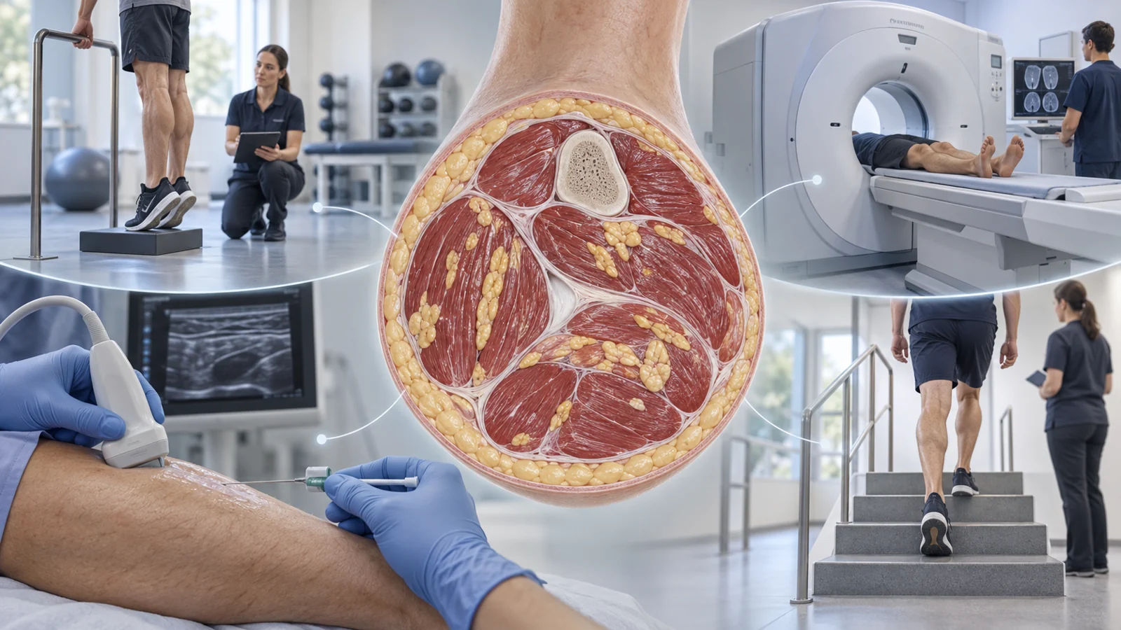
A planned study called IMAT Quality in Aging is asking whether calf strengthening might change more than muscle force. It will examine fat within the muscle, known as intramuscular adipose tissue or IMAT, alongside muscle composition, tissue markers, and everyday movement measures.
The study’s central hypothesis is that IMAT in older muscle may show more damage and pro-inflammatory signaling with aging, and that progressive resistance exercise may reduce those features. The supplied registry record describes this as planned research, not a completed finding. It lists the study as NOT_YET_RECRUITING, with an estimated start date of October 1, 2026, and estimated primary and overall completion in January 2028. No results are posted in the record used here.
The relevant question is what the planned protocol is designed to distinguish: a change in how much muscle or fat is present, a change in the biological activity of sampled tissue, and a change in how well the leg performs.
The question behind the calf intervention
The study is focused on 16 adults aged 65 to 80 who are described as underactive. Researchers will compare measurements taken before and after a 12-week calf resistance program. Each participant supplies their own baseline, allowing the investigators to ask whether several features of the same person’s calf look different after the intervention.
The exercise is delivered in small groups over Zoom with a trained therapist, three times per week. The protocol specifies standing heel rises, seated heel rises, and long-sitting ankle plantarflexion. Resistance may come from body weight, a weighted vest, or a resistance band, with loading progressed as strength improves. These details describe the study exposure; they are not a recommendation for an individual exercise program.
Participation also includes screening for eligibility and safety, an initial assessment of calf strength and physical performance, a CT scan of the lower leg, and an ultrasound-guided biopsy of the gastrocnemius muscle and nearby IMAT. About a week after the program ends, participants return for repeat strength testing, CT imaging, and biopsy. The planned follow-up is approximately 14 weeks after baseline.
Two ways of looking at the calf
CT and biopsy answer different questions. CT is used to estimate the volume of the plantarflexor muscle compartment and the volume of fat within that compartment. It provides a structural view: how much muscle and internal fat occupy the sampled region after image processing separates them from bone and subcutaneous fat.
A biopsy provides a much closer look at tissue biology. The study’s primary outcome is senescent-cell density, measured in histological sections using β-galactosidase and p16 expression. Investigators will count positive cells and normalize the count to the tissue area examined. This is a planned marker-based measurement. It is not evidence that the exercise program clears senescent cells or functions as a senolytic treatment.
The distinction matters because a change in fat volume would not automatically describe what the fat or surrounding cells are doing. Conversely, a change in cellular markers could occur without a large visible change in the total amount of fat. The protocol is designed to collect both kinds of information rather than treating one measurement as a substitute for the other.
From tissue markers to secreted signals
The biopsy work extends beyond counting cells. The registry describes a sequence that begins with bulk RNA sequencing to identify genes whose expression may respond to age-related biology or exercise and that encode secreted proteins. Candidate signals would then be checked by measuring proteins released from cultured biopsy tissue.
If signals are validated, RNA in situ hybridization, including RNAscope, is planned to help locate their cellular origin within the tissue. Histological sections will also be used to examine adipocyte size, macrophage density, senescent cells, and beige adipocytes. Together, these tests are intended to connect a molecular candidate with a measurable protein and then with the cells and locations where it appears.
That sequence remains prospective. The study has not yet supplied evidence that a particular signal is elevated, reduced, inflammatory, or exercise-responsive in participants. It describes how researchers plan to move from gene-expression candidates to protein measurements and spatial tissue information.
Movement tests add a practical measure
The study does not stop at the microscope. Researchers will measure ankle plantarflexor peak torque, stair-climb power, walking speed, and a broader physical performance score. The score combines tasks such as standing balance, repeated chair rises, turning, picking up an object, walking, and stair performance.
These tests represent different aspects of lower-limb function. Ankle torque measures the turning effect of muscle force at the ankle during a controlled test. Stair-climb power is calculated from body weight, the vertical height climbed, and the time taken. Walking speed captures movement over a short course, while the broader score combines balance, coordination, strength, and task completion.
Pairing these outcomes with CT and biopsy allows the researchers to ask whether tissue-level changes and functional changes appear together. It would still take additional evidence to show that one caused the other. A person might perform differently for reasons not captured by the tissue measurements, and a biological change might not be large or relevant enough to alter movement.
What the planned design can show
A 16-person, single-group before-and-after study can provide detailed measurements in a small, defined population, but it has important limits. Participants are compared with their own baseline rather than with a parallel untreated group. Any eventual difference over the study period would therefore need cautious interpretation, because the design alone cannot separate an exercise effect from every other change occurring over time.
The cohort is also restricted to older adults described as underactive, and the tissue sample comes from the calf. The study can describe what happens in this selected group and sampled region. It cannot, by itself, establish how the starting tissue state differs from that of younger adults, because the supplied record does not establish an additional age-comparison dataset.
The registry’s estimated dates also describe plans rather than verified events. The record lists an estimated October 2026 start and estimated January 2028 completion, while its current status is not yet recruiting. Until the study generates and posts results, the proposed reductions in inflammatory signaling, changes in IMAT volume, gains in strength, and improvements in mobility remain questions rather than findings.
What this could mean for mobility in older age
The longer-term possibility is practical. If the study identifies reproducible biological responses in fat within aging muscle, and if those responses are connected to meaningful improvements in strength, walking, or stair-climbing ability, muscle-fat biology could eventually help inform resistance programs intended to preserve mobility in older adults.
That pathway requires more than a promising tissue marker. It depends on repeatable results, functional benefit, and stronger evidence that the biological changes are linked to how people move. At this planned-study stage, the potential value lies in testing that connection carefully—not in claiming that the registered program has already delivered it.
Key Sources
- Primary source: IMAT Quality in Aging
Government or Regulator · Clinical Trial · 2026-04-02
Keep exploring