
Research Signal – Source date 2025-04-01
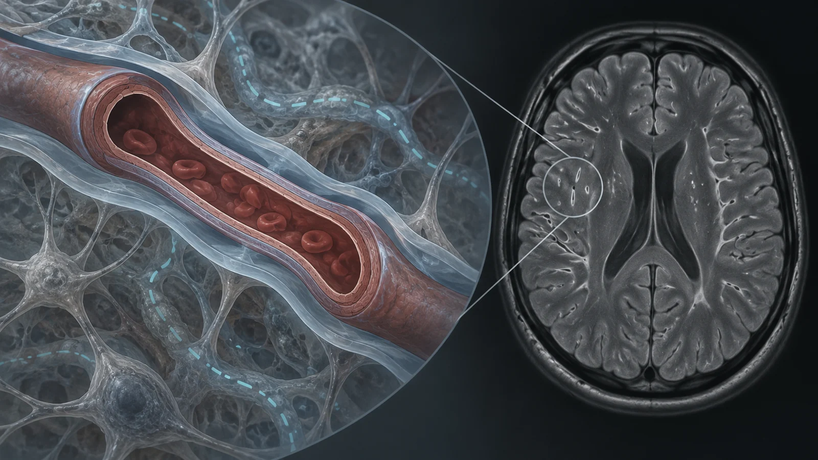
Cerebral small vessel disease affects the brain’s smallest blood vessels and is a major vascular contributor to cognitive impairment and dementia. On MRI, one feature associated with the disease is a greater number of visible perivascular spaces: fluid-filled channels that surround blood vessels.
Could those spaces show that the brain is struggling to clear waste? They may offer a clue, but a scan of an enlarged space cannot answer that question by itself. A review published in April 2025 surveyed English-language human and rodent studies published through December 2024. It describes a fast-developing set of imaging approaches, while emphasizing that none of the proposed non-invasive measures has been formally validated as a direct test of waste clearance involving these spaces.
The fluid route is still under debate
Perivascular spaces, or PVS, are channels around vessels that may participate in exchanges between cerebrospinal fluid (CSF) and interstitial fluid (ISF), the fluid between brain cells. One proposed account holds that CSF enters along small arteries, exchanges fluid and dissolved substances with the brain’s tissue, then leaves through routes near veins and through lymphatic vessels around the brain. In this view, arterial pulsation and water channels called aquaporin-4 may help shape movement.
Other accounts differ on how fluid and substances leave the brain. The intramural periarterial drainage hypothesis proposes outflow along basement membranes within artery walls, driven in part by the motion of vascular smooth muscle. A “mixing” account instead places bulk flow mainly at the brain’s surface, where CSF and ISF mix before moving toward lymphatic vessels.
The direction and driving force of fluid movement remain contested. Some studies support bulk flow along vessels; others suggest that diffusion—the spreading of molecules—may explain much of the exchange. The review presents these as competing, and potentially complementary, explanations rather than a settled map of how fluid moves in people with small vessel disease.
What a routine MRI can show
Most perivascular spaces are too small to see on conventional MRI. The ones that are visible appear as fluid-like channels; when imaged perpendicular to a vessel, the standard definition cited by the review describes them as less than 2 millimeters across. Their visibility makes them countable, but does not reveal what is happening in every surrounding channel or directly measure clearance.
Sequence choice affects what radiologists see. T2-weighted MRI is preferred for identifying visible PVS. T1-weighted scans can also be used, though the review notes that rating from T1 alone may underestimate their visibility. Calcified vessels and other tissue changes can complicate interpretation. Automated approaches may quantify their number, shape or volume, but algorithms can confuse PVS with lesions such as small cavities or white-matter changes, so their output still needs visual review.
Visible PVS are recognized among the imaging markers of small vessel disease and may appear before some other markers, including small areas of tissue damage and white-matter changes. A higher visible PVS burden has been associated with disease burden and cognitive impairment in studies covered by the review. Researchers still do not know exactly why spaces become enlarged and visible. Tissue loss may contribute; impaired fluid circulation, protein or immune-cell accumulation, and blood-vessel changes have also been proposed.
Different scans capture different pieces
Researchers have tried several ways to look beyond visible spaces. Dynamic contrast-enhanced MRI after gadolinium is injected into the fluid around the brain can track contrast as it moves through CSF and into parts of the brain. In one human study, researchers compared this approach with DTI-ALPS, a measure calculated from diffusion MRI. The comparison found a relationship between the two measures, but they sampled different regions: DTI-ALPS used a small area of white matter, while the contrast measurements included mainly ventricular and cortical areas. Agreement between the measures therefore does not show that they capture the same process in the same place.
DTI-ALPS estimates the direction of water diffusion in a small white-matter region. Lower values have been associated with greater small vessel disease burden and cognitive impairment. But the method uses signals from millimeter-scale MRI voxels, too coarse to isolate the much smaller PVS themselves. It is a non-invasive proxy whose connection to waste clearance remains unvalidated as a direct measurement.
Contrast-enhanced imaging offers a different kind of evidence: it follows a tracer, but requires an injection into the fluid surrounding the brain, repeated scans and a contrast agent the review describes as potentially harmful. Those demands limit its use. Other methods, including arterial spin labeling, estimate blood flow by magnetically labeling blood. They could offer indirect information about fluid dynamics, but blood flow is not itself a direct measurement of CSF–ISF exchange, and the proposed link remains speculative.
These techniques observe distinct things: visible spaces, tracer movement, water diffusion or blood flow. Comparing them can help researchers understand what each might reveal. It cannot yet turn any one of them into a validated clearance test.
Baseline associations leave a timing question
A scan taken at one point in time can be associated with what happens later without showing how the measured feature changes along the way. In the limited longitudinal studies the review discusses, baseline measures—visible PVS volume and/or DTI-ALPS—were related to later progression of small vessel disease and cognitive impairment.
But changes in those imaging measures over time did not correlate with future cognitive decline after accounting for white-matter integrity. That finding complicates a simple prediction story: a baseline association is not the same as evidence that worsening scan measures track worsening cognition. More longitudinal research is needed to establish how these measures change and what those changes mean.
A possible feedback loop, still untested over time
The review describes factors that could connect small vessel disease with disrupted fluid exchange. Aging can bring changes to small vessels and reduced arterial pulsation; hypertension can stiffen vessels and impair blood-flow regulation. Neuroinflammation has been associated with disruption of the blood–brain barrier (BBB), which regulates movement between blood and brain, and may also interfere with fluid entry into PVS.
The BBB and waste clearance involving PVS may also affect one another. If either pathway handles amyloid beta less effectively, the other could be affected, potentially encouraging further accumulation. The review presents this as a proposed reinforcing cycle, not a sequence demonstrated in people over time. Human longitudinal evidence establishing how barrier changes and clearance changes influence each other remains absent from the research it discusses.
What better brain scans could mean for small vessel disease
If researchers validate imaging measures against the underlying fluid processes and show how they relate to disease over time, scans could eventually help identify earlier brain-fluid changes or track disease severity and response to treatment. For people with small vessel disease, that could make it easier to distinguish meaningful progression from a single ambiguous scan finding.
That possibility depends on resolving what each method measures, establishing reliable reference values and linking changes to outcomes in longitudinal studies. The review does not establish that these measures can guide treatment today. Their eventual clinical value depends on a clearer account of fluid movement and validated links between scan findings and the course of disease.
Key Sources
- Primary source: Imaging brain fluid dynamics and waste clearance involving perivascular spaces in cerebral small vessel disease.
DOI or Journal Page · Review or Meta-Analysis · 2025-04-01
Keep exploring