
Research Signal – Source date 2023-03-17
A head injury is easy to understand as an event: an impact, a period of acute treatment, and an expected recovery. The harder question is what may happen after that immediate phase. Some people experience persistent changes in memory, concentration, mood, or everyday function, and research has also linked traumatic brain injury with neurodegenerative changes that can emerge over much longer timescales.
A March 17, 2023 mini-review, Inflammaging, cellular senescence, and cognitive aging after traumatic brain injury, examines one possible explanation. It brings together epidemiological and clinical observations, experimental brain-injury studies, and post-mortem findings to ask whether injury can push some brain cells into a persistent inflammatory state. The proposal is not that every later cognitive problem has one cause. It is that the original injury may set biological processes in motion that continue after the first damage has passed.
The long tail after the original injury
The review describes traumatic brain injury as a possible starting point for progressive neurodegeneration, whether after one moderate-to-severe injury or after repeated mild injuries. In the evidence it discusses, chronic inflammation is associated with white-matter damage and cognitive dysfunction years after injury. White matter helps connect brain regions, and its deterioration can accompany problems with memory, concentration, mood, and other aspects of mental function.
This long time horizon changes the scientific question. Acute injury research often focuses on axonal damage, neuronal loss, swelling, and pressure inside the skull. Those processes matter, but they do not by themselves explain why tissue changes and cognitive problems may continue long after the initial event. The review therefore considers whether aging-related biology could become entangled with the aftermath of brain trauma.
One concept at the center of that possibility is inflammaging, a low-level, persistent inflammatory state associated with natural aging. Inflammation is useful in responding to injury and infection. When immune activity remains active or becomes poorly regulated, however, it can also contribute to chronic tissue stress. The proposed connection is that traumatic brain injury may accelerate some of the cellular changes normally associated with aging, creating a route toward premature cognitive decline.
How a damaged glial cell could keep the process going
Cellular senescence is a state in which a cell stops dividing but remains alive and metabolically active. It is not simply a cell that has died or gone quiet. Senescence can follow several kinds of stress, including DNA damage, oxidative stress, mechanical stress, disrupted autophagy, and inflammatory signals. The review highlights oxidative and genotoxic stress after acute brain injury as possible triggers of senescence pathways.
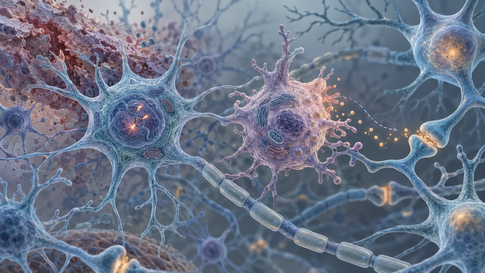
The brain-support cells called glia are especially relevant here. Glia include cells that help maintain the brain’s environment, respond to injury, support nerve fibers, and participate in immune defense. Unlike mature neurons, which do not undergo senescence in the same way, glial cells can show senescence-associated changes. After experimental brain injury, researchers have observed increased markers associated with senescence in areas including the cortex, hippocampus, and thalamus. The review also describes senescence-associated findings in microglia and astrocytes, two important glial populations.
A senescent cell can also develop what researchers call a senescence-associated secretory phenotype, or SASP. In plain language, its behavior changes so that it releases signaling molecules such as inflammatory cytokines and chemokines. These signals can attract immune cells, influence neighboring cells, and alter the surrounding tissue. Matrix-remodeling enzymes released in this state may also affect the tissue scaffold around cells.
This gives the review a proposed sequence rather than a settled diagnosis. Injury-related stress may damage DNA and activate senescence pathways. Senescent glia may then remain present and release inflammatory signals. Those signals could help sustain inflammation, alter repair, and add stress to nearby neural connections. Over time, the interaction between damaged tissue and persistent immune activity might contribute to worsening brain structure and function.
The sequence is biologically plausible, but the supplied evidence does not establish that senescent cells cause delayed cognitive deterioration in people after traumatic brain injury. The value of the proposal is that it connects a short-lived event with a potentially persistent cellular state that can be tested.
What human tissue and animal studies each show
The evidence comes from settings that answer different questions. In post-mortem brain tissue from contact athletes with histories of brain trauma, researchers observed glial senescence-associated features and SASP-like phenotypes. Those observations make the proposed biology relevant to human neurotrauma, but tissue examined after death cannot by itself show when the changes began, how they evolved, or whether they caused the person’s symptoms.
Experimental studies offer a different kind of information. In rodents exposed to moderate or repeated mild brain injury, senescence-associated beta-galactosidase and other senescence-related markers increased in injured brain regions. Some findings persisted beyond the immediate injury period. These experiments allow researchers to examine timing and tissue responses more directly, but a marker is not the same as proof that senescent cells are driving later cognitive change.
The review also discusses research in Alzheimer’s disease and tau-related neurodegeneration models. In those models, removing or reducing senescent cells was associated with less inflammation, less pathological protein accumulation, and better cognitive outcomes. Such findings help show why senescence is being considered as a therapeutic target in neurological disease. They remain separate from traumatic brain injury evidence, however. A result in an Alzheimer’s or tauopathy model does not establish an equivalent effect after head trauma.
One brain-injury example is particularly informative because it complicates any simple treatment story. In mice exposed to repetitive mild traumatic brain injury, the experimental drug navitoclax reduced senescence markers and improved cognitive function in male mice. The corresponding benefit was not observed in female mice. This sex-dependent result suggests that the biology may vary across groups, but it does not establish how men and women would respond to treatment. Nor does one mouse experiment establish a safe or effective human intervention.
Why removing a cell is different from quieting its signals
The proposed treatments fall into two broad categories. Senolytics are intended to remove senescent cells. Senomorphics are intended to change what those cells do, including reducing harmful inflammatory signaling without necessarily killing the cells. The distinction matters because a cell can influence its surroundings through its secretions, and removing the cell is not the only way to alter that influence.
The review describes several experimental possibilities, including fisetin, navitoclax, curcumin-related compounds, and other approaches that may affect senescence, inflammation, metabolism, or cell survival. Many of these substances have multiple biological actions. If a compound reduces inflammation or improves cognition, that result does not by itself show that selective removal of senescent cells caused the benefit. The same caution applies to the review’s discussion of curcuminoid supplementation in critically ill patients with traumatic brain injury. Those clinical findings concern a particular patient population and reported outcomes; they are not proof of selective senescent-cell clearance or prevention of delayed cognitive decline.
The practical challenge is to identify which cells should be targeted, when they should be targeted, and which aspects of their activity are harmful. Senescent-like changes may not be identical across cell types or stages of injury. A treatment that removes cells broadly could affect functions that remain useful, while a treatment that only dampens inflammatory signals might leave other damaging processes untouched. The supplied evidence does not authorize a patient regimen or an optimal treatment window.
The immune response cannot simply be switched off
Inflammation is one reason this treatment direction requires care. Immune activity can contribute to defense and repair after injury, even while prolonged activation is associated with later damage. A broad anti-inflammatory intervention could therefore suppress helpful responses along with harmful ones.
The review points to the large CRASH corticosteroid trial as a warning against assuming that general immune suppression will improve outcomes after brain injury. The trial was stopped early because outcomes were worse in the corticosteroid arm than in the placebo arm. That result is not a direct test of senolytic treatment, and it does not answer whether a precisely targeted senomorphic or senolytic approach could help. It does show why the therapeutic goal cannot be reduced to “less inflammation” in every circumstance.
A more specific strategy would need to preserve useful immune activity while limiting the persistent signals that damage tissue or interfere with recovery. That requires evidence linking a target to a particular stage of post-traumatic biology, along with safety testing and durable outcome measures. Cognitive improvement in an animal experiment is an important result, but it is only one part of that chain.
What targeting senescent cells could mean for life after brain injury
For people living with the long-term consequences of traumatic brain injury, the attraction of this idea is practical. A treatment that could limit continuing cellular and inflammatory damage might help preserve memory, everyday function, and quality of life beyond the initial recovery period. It could shift care from responding only to the original injury toward protecting the brain during the years that follow.
That possibility remains conditional. Researchers would first need to establish a causal, safely targetable role for senescent cells after human brain injury and then demonstrate a durable benefit in people. The 2023 review provides a reason to investigate that route: injured brain-support cells may help connect an early physical insult with persistent inflammation and later cognitive deterioration. It does not yet turn that proposed connection into an established treatment.
Key Sources
- Primary source: Inflammaging, cellular senescence, and cognitive aging after traumatic brain injury.
DOI or Journal Page · Review or Meta-Analysis · 2023-03-17
Keep exploring