
Research Signal – Source date 2026-02-17
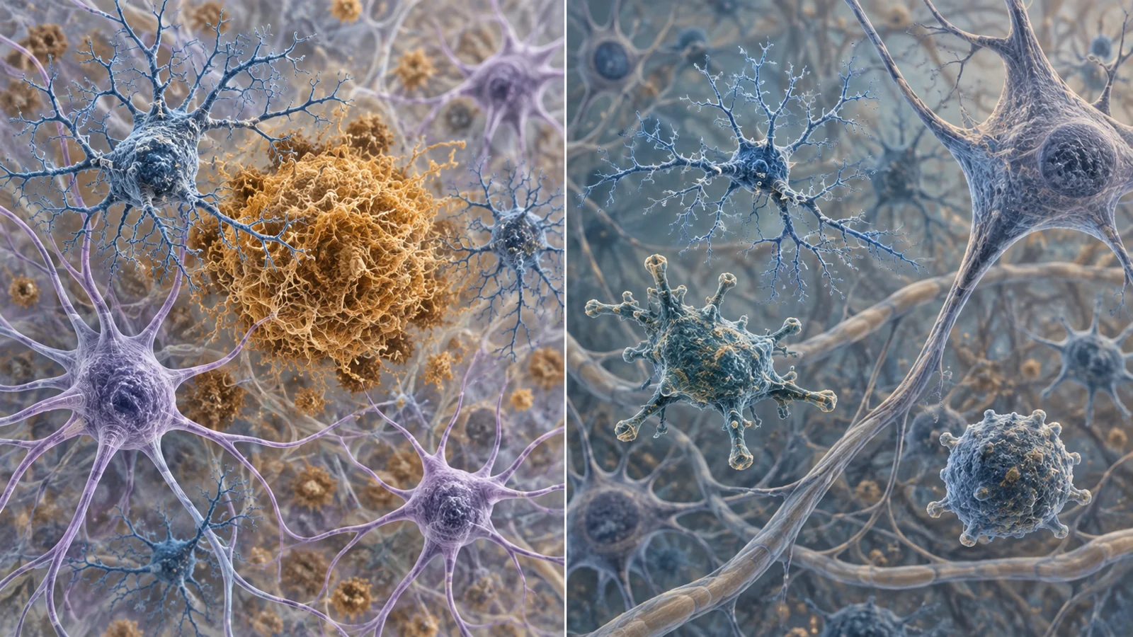
Microglia are the brain’s resident immune cells. Astrocytes help support neurons and their connections, and can change their behavior when the brain is stressed or damaged. Both cell types respond to trouble, but “activated” does not name one uniform state or tell us whether a cell is helping, harming, or doing both.
A February 17, 2026 review compares disease-associated microglia and astrocytes across developmental stress, neurodegeneration and cellular senescence. Its examples raise a practical question: when studies report brain inflammation, what are these cells responding to, and why might their roles differ between Alzheimer’s disease and amyotrophic lateral sclerosis (ALS)? The answer depends on which cells are present, where and when they change, and how the evidence was gathered.
From damaged material to a changing immune response
When neural cells are injured or die, they can release host-derived molecules that act as danger signals. Microglia recognize such signals through receptors involved in innate immunity. In neurodegenerative settings, the review describes signals from damaged neural material, myelin debris, lipid breakdown products and protein aggregates as possible prompts for disease-associated microglial responses.
One proposed role for these responses is to contain and clear damage. That can be useful in principle, but the consequences depend on the surrounding tissue and the state the cells enter. A response directed at debris or aggregates takes place within a living network of neurons and other cells. If the response changes over time or recruits inflammatory processes, its effects may differ from those of an early attempt to contain injury. The label “disease-associated” describes a profile observed in a disease context; it does not, on its own, establish that the profile causes disease or is uniformly damaging.
Alzheimer’s disease has local and shifting glial states
In Alzheimer’s disease (AD), the review describes disease-associated astrocytes near amyloid-beta plaques and disease-associated microglia. The astrocyte state appears early in the reviewed studies and expands as AD progresses. Reports from mouse and human material also describe similar astrocyte states with aging, including cells near plaques that express inflammatory markers such as SerpinA3N. These findings make location and timing part of the story: a cell’s profile is observed in relation to nearby pathology and to a particular stage, rather than as a whole-brain switch.
Microglial profiles in AD also resist a simple good-or-bad division. A study summarized by the review identified pro-inflammatory and anti-inflammatory DAM subtypes in mouse models. In human datasets, pro-inflammatory markers were associated with neuropathology and appeared before cognitive decline. In the mouse-model work, compounds that shifted profiles toward an anti-inflammatory pattern were also associated with greater amyloid clearance. The human association and the mouse intervention answer different questions: one links markers with disease features, while the other tests a manipulation in an experimental model. Neither establishes that shifting the same profile would improve outcomes in people.
The proximity of astrocytes and microglia to plaques is informative about where these cells are found and how their states relate to local disease features. It does not by itself reveal whether a cell state initiates damage, responds to it, or changes the course of disease. Those possibilities require evidence beyond a cell’s location or molecular signature.
ALS shows why the same label can conceal a different mix
The review’s ALS examples do not reproduce the AD pattern. In ALS tissue, elevated expression of genes including TREM2, MS4A, CD33, APOE and TYROBP has been observed in postmortem spinal cord samples. But a disease-associated label does not mean the same composition in each condition. The review contrasts a phagocytic subtype prominent in AD with an inflammatory subtype prominent in ALS in the studies it discusses.
Human profiling adds another complication. A study of ALS motor cortex and spinal cord tissue classified microglia into roughly seven subtypes. The most prevalent group had an undifferentiated profile with disrupted respiratory electron-transport signatures, while the DAM-like subset was reduced. The authors of that work proposed that a reduction in this subset might characterize later disease. This is a tissue observation and interpretation, not evidence that restoring DAM-like cells would help patients.
A separate experimental route comes from induced astrocytes made from ALS patients. Those cells showed neurotoxic effects in culture, and their measured features varied among patients. In that cell-culture work, restoring miR-146a in depleted astrocytes reversed an inflammatory state. The result suggests that patient-derived cells can be used to investigate different mechanisms, but it does not merge with the tissue findings or demonstrate a treatment effect in people with ALS.
Stress can travel through several cell types
Developmental and social stress examples show how a glial response may involve a wider circuit of cells. In one mouse model combining maternal infection with social stress, researchers identified a stress-associated microglial state in the cerebellum, alongside Purkinje neuron loss. Replacing microglia systemically or in the cerebellum improved cerebellar dysfunctions in that model. Aged human samples also contained cells with a similar stress-associated profile, but that observation does not establish the same sequence or effect in people.
Another mouse study traced a route from astrocytes to neutrophils. Astrocyte-derived CCL5 signaling was linked to CCR5-positive neutrophils entering the hippocampus, neuronal damage and depression-like behaviors. Interventions involving bone marrow cells or inhibition of the CCL5/CCR5 pathway alleviated those findings in the model. This example is not simply a story about microglia switching state: it describes communication between astrocytes and immune cells that can enter brain tissue. Its behavioral and intervention results remain specific to the experimental setting.
Aging changes what a senolytic result can mean
Senescence adds an age-dependent boundary to the idea that removing harmful cells will have a consistent effect. In a mouse model of demyelination, senescent-like microglia and macrophages accumulated in lesions with age. The review describes a senescence-associated secretory profile that included CCL11, which impeded oligodendrocyte maturation. Senolytic treatment restored myelin regeneration in younger mice, but not in aged mice.
That difference matters for interpretation. The experiment supports an age-dependent result in this demyelination model; it does not show that senolytics broadly clear senescent brain cells or restore function in older people. Age may change the cellular environment in which an intervention acts, so an effect in younger animals cannot simply be carried over to older ones.
Cell profiles and imaging answer different questions
Single-cell and single-nucleus profiling can separate cell populations and compare their gene or protein signatures across regions and conditions. The human microglia studies summarized in the review distinguish subgroups associated with processes such as antigen presentation, movement and proliferation. Such measurements help describe which states are present in the sampled tissue. They do not alone show what those cells do over time or whether a particular state drives symptoms.
Imaging offers a different kind of view. The review discusses monoamine oxidase B (MAO-B), an enzyme with relatively high astrocyte specificity, as a target for positron emission tomography (PET) tracers. Imaging with these tracers can help observe astrocyte-related changes in a living brain, including around dense amyloid plaques. A signal is still a marker: its interpretation depends on tracer properties and does not by itself establish the function or cause of the labeled cell state.
Both approaches also depend on what tissue or signal is available. Human postmortem studies face limits related to access, the interval between death and tissue collection, and the regions that can be sampled. Animal models and cell systems allow different kinds of experiments, but their results need to be interpreted in their own settings. Reading across methods can sharpen the questions; it cannot turn associations, profiles and interventions into one shared measure of cause or treatment effect.
What better maps of brain inflammation could make possible
More precise ways to distinguish glial states could eventually help researchers identify which brain processes are worth targeting in a particular disease stage or age group. If cell profiles, spatial measurements and living-brain imaging can be linked to meaningful changes in function, they may support better choices about which mechanisms to test and which patients or models are most relevant. That could make future treatments more focused than a broad attempt to suppress brain inflammation.
The path to that benefit remains conditional. The review gathers selected mouse, human-tissue, cell-culture and imaging studies, and it does not establish that changing a glial state improves outcomes in people. Showing which states cause harm, which are protective, and whether those roles shift with disease stage or age remains essential before a cell profile can guide treatment.
Key Sources
- Primary source: Glioinflammation: disease-associated microglia and astrocytes in psychiatric disorders, neurodegeneration, and senescence.
DOI or Journal Page · Review or Meta-Analysis · 2026-02-17
Keep exploring