
Research Signal – Source date 2023-01-02
The brain is not an isolated mass of cells. It is surrounded and penetrated by moving fluids that help distribute substances, respond to pressure and transport material through several connected spaces. Researchers are investigating whether changes in these fluid movements could help explain why aging is associated with a higher risk of dementia, including Alzheimer’s disease.
A review published on January 2, 2023, brings together animal experiments, human imaging observations and findings from human brain tissue. Its contribution is not the announcement of a new treatment or a settled mechanism. Instead, it connects three questions that are often discussed separately: where fluid travels, how brain cells and blood vessels influence that movement, and what current measurements can actually establish about dementia.
A proposed circulation system around brain cells
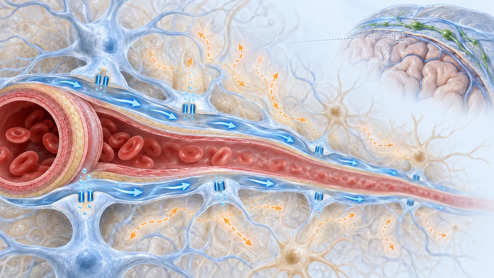
Cerebrospinal fluid, or CSF, occupies spaces around the brain and spinal cord. Some of it travels through the subarachnoid space, the fluid-filled region between protective layers surrounding the brain. Research summarized in the review shows a preferential route along spaces beside arteries. These vessel-adjacent pathways can carry fluid toward brain tissue and are therefore central to the idea that CSF participates in moving substances through the brain.
The glymphatic system is the name given to a proposed brain waste-clearance arrangement involving these perivascular spaces, the fluid between brain cells and channels beside veins. In the hypothesis, CSF enters along artery-adjacent routes, exchanges with interstitial fluid in the brain’s tissue and helps carry dissolved material toward an exit pathway. Drainage may then involve spaces near veins and meningeal lymphatic vessels in the brain’s dural covering. Communication routes between the skull and brain are also part of the wider picture described by the review.
The physical details matter because “clearance” can describe more than one kind of movement. Diffusion is the spreading of molecules down concentration differences. Bulk flow, sometimes called advective flow, is movement in which fluid carries dissolved material along. Smaller molecules moving over shorter distances may be influenced strongly by diffusion, while bulk movement may contribute over longer anatomical distances. The review presents fluid dynamics as a combination of processes that varies with the brain compartment, the material being moved, age, sleep and health.
Evidence for one part of this map is stronger than evidence for the whole proposed circuit. Experiments in animals and observations in people show CSF moving preferentially along periarterial spaces. But whether the movement continues as a single, one-way flow through brain tissue remains unsettled. The review also states that direct clinical or preclinical evidence for CSF crossing from brain tissue into perivenous channels is lacking from its January 2023 perspective.
Why the brain’s movement is not one simple flush
The glymphatic hypothesis became influential because it offered an intuitive picture of how hydrophilic waste products, which do not readily cross the blood-brain barrier, might leave brain tissue. Yet the proposed transport mechanism has been challenged by both experiments and physical analysis.
One issue is direction. Tracers introduced into fluid spaces can travel along periarterial routes before entering brain tissue. In the opposite direction, other experiments have found that tracers placed directly into brain tissue can later accumulate around arteries. Those observations leave open the possibility that perivascular movement is bidirectional rather than a single stream entering by arteries and leaving by veins.
Another issue is the amount of flow required. The resistance to fluid movement through the narrow extracellular spaces between cells may make large-scale advective transport difficult. Computational analysis discussed in the review suggests that the flow needed to clear large molecules at some proposed rates could exceed the amount of CSF produced in the brain. A different interpretation is that CSF in perivascular spaces acts as a sink, allowing solutes to diffuse toward it rather than being carried through the tissue by a continuous bulk current.
These competing explanations do not make the observed movement irrelevant. They change what it means. Researchers can observe routes, timing and changes in tracer distribution without having proved that the brain operates a complete, one-way cleansing circuit. For dementia research, that distinction separates a plausible physical connection from a demonstrated cause of disease.
Arterial motion, breathing and sleep change the exchange
Fluid movement is not driven by a single pump. The review describes arterial pulsation, changes in vessel diameter called vasomotion and respiration as influences that can create pressure differences and alter exchange between CSF and interstitial fluid.
A mouse experiment illustrates the importance of identifying the physical driver. When researchers ligated an internal carotid artery to reduce arterial pulsatility, the animals showed reduced exchange between CSF and interstitial fluid. The result supports the idea that the motion of blood vessels can help shape fluid transport. It does not, by itself, show that the same intervention or relationship determines dementia in people.
Sleep is another physiological state associated with differences in fluid exchange. The reviewed research reports greater CSF-interstitial fluid exchange during sleep and changes in the movement of intrathecal contrast depending on sleep or wakefulness. This makes sleep relevant to the biology being measured, while leaving open how much of the relationship is shared across species, brain regions and disease states. It is a reason to study brain physiology carefully, not a prescription for preventing dementia.
Aquaporin-4 makes location as important as quantity
Aquaporin-4, or AQP4, is a water-channel protein found on the endfeet of astrocytes. Astrocytes are support cells whose extensions contact blood vessels and help form the boundary between vessel-adjacent spaces and brain tissue. Because AQP4 sits at this interface, researchers have tested whether it helps regulate the exchange of fluid and solutes.
Animal experiments have produced evidence in both directions. Some studies summarized by the review found that reducing or removing AQP4 lowered tracer clearance, while other studies did not reproduce a change in tracer movement after AQP4 deletion. The disagreement matters because it means the proposed role of the channel cannot be treated as a settled switch that simply turns clearance on or off.
Human tissue findings add a different layer. The review reports increased AQP4 expression in the aging human brain, while reduced localization around blood vessels is associated with Alzheimer’s pathology. Reduced perivascular localization is associated with greater Alzheimer’s-related neurofibrillary and amyloid-beta pathology, even after accounting for age in the analysis described by the review.
The distinction between amount and placement is important. More of a water-channel protein overall does not necessarily mean that it is positioned where fluid exchange requires it. At the same time, an association between altered localization and Alzheimer’s pathology does not establish which came first. The animal manipulation studies test a mechanism under experimental conditions; the human tissue observations show a relationship in disease-associated brains. Together they identify a biological question, not a proven causal chain from mislocalized AQP4 to dementia.
Human scans show routes more readily than clearance
Researchers need ways to study fluid movement in people without disrupting the system they are trying to understand. One established approach has used intrathecal gadolinium contrast, followed by repeated imaging as the agent moves from the lumbar fluid space toward the brain. These studies show rapid transport into basal cisterns, over the cerebral surface and along large arterial pathways before contrast enters brain tissue.
Such images are useful because they reveal where a substance appears and how its distribution changes over time. They support important parts of the periarterial transport model and show that sleep and cardiopulmonary motion can influence what is observed. But the spatial resolution of MRI and PET does not settle what happens inside the smallest penetrating perivascular spaces. Contrast arrival also does not directly demonstrate bulk flow through brain tissue or prove that a measured signal represents the removal of harmful metabolic waste.
Newer approaches aim to reduce those limitations. The review discusses diffusion-weighted MRI as a possible way to quantify movement in perivascular spaces without gadolinium contrast. Other studies have used changes in intravenously delivered gadolinium within brain tissue as an indirect marker of clearance. These approaches may make repeated or less disruptive measurement more practical, but they remain indirect measures that require validation against the underlying biology.
The central measurement problem is therefore two-sided. Injected tracers can make movement visible, yet introducing a substance or altering a barrier may disturb normal physiology. Non-invasive signals may preserve more of the natural system, yet a signal that changes with fluid motion is not automatically a validated measure of waste removal. Better quantitative imaging is needed to connect observed transport with the disease processes researchers want to understand.
What understanding brain clearance could mean for dementia care
The potential value of this research is a clearer biological process to target if impaired fluid exchange contributes to dementia. Preserving or restoring useful clearance could eventually become one route toward protecting brain function, but that possibility depends on establishing a causal contribution in humans and showing that the process can be changed safely in ways that improve meaningful outcomes for patients.
The January 2023 review does not establish that reduced clearance causes Alzheimer’s disease or that boosting it prevents, treats or reverses dementia. It does show why the question remains scientifically important. Fluid routes, vessel motion, sleep-related state changes and the placement of AQP4 may connect the brain’s physical environment with the accumulation of disease-associated pathology. The next advance would be more than a brighter scan or a more persuasive diagram: it would be reliable evidence linking a measurable fluid process to human disease and to a safe, useful clinical intervention.
Key Sources
- Primary source: Fluid dynamics in aging-related dementias.
DOI or Journal Page · Review or Meta-Analysis · 2023-01-02
Keep exploring