
Research Signal – Source date 2021-07-09
Disturbed sleep and dementia often appear together, but that association leaves an important question open. Is the connection happening only through disrupted rest and daily behavior, or does altered timing reach into the machinery of brain cells themselves?
A review published on July 9, 2021, examines that question through circadian clock genes, the molecular system that helps cells organize activity across the day. It is not a new treatment experiment. Instead, it brings together observations from people, animal models, and cellular studies to explain how daily timing might intersect with processes involved in neuronal injury and cognitive loss.
The review’s central idea is more specific than “better sleep protects the brain.” It asks whether clock-linked gene activity helps coordinate waste handling, cellular recycling, energy regulation, and neuronal survival. The evidence points toward a connected biology, while the distance between these mechanisms and a treatment that preserves memory in people remains substantial.
A daily clock is a gene-control system
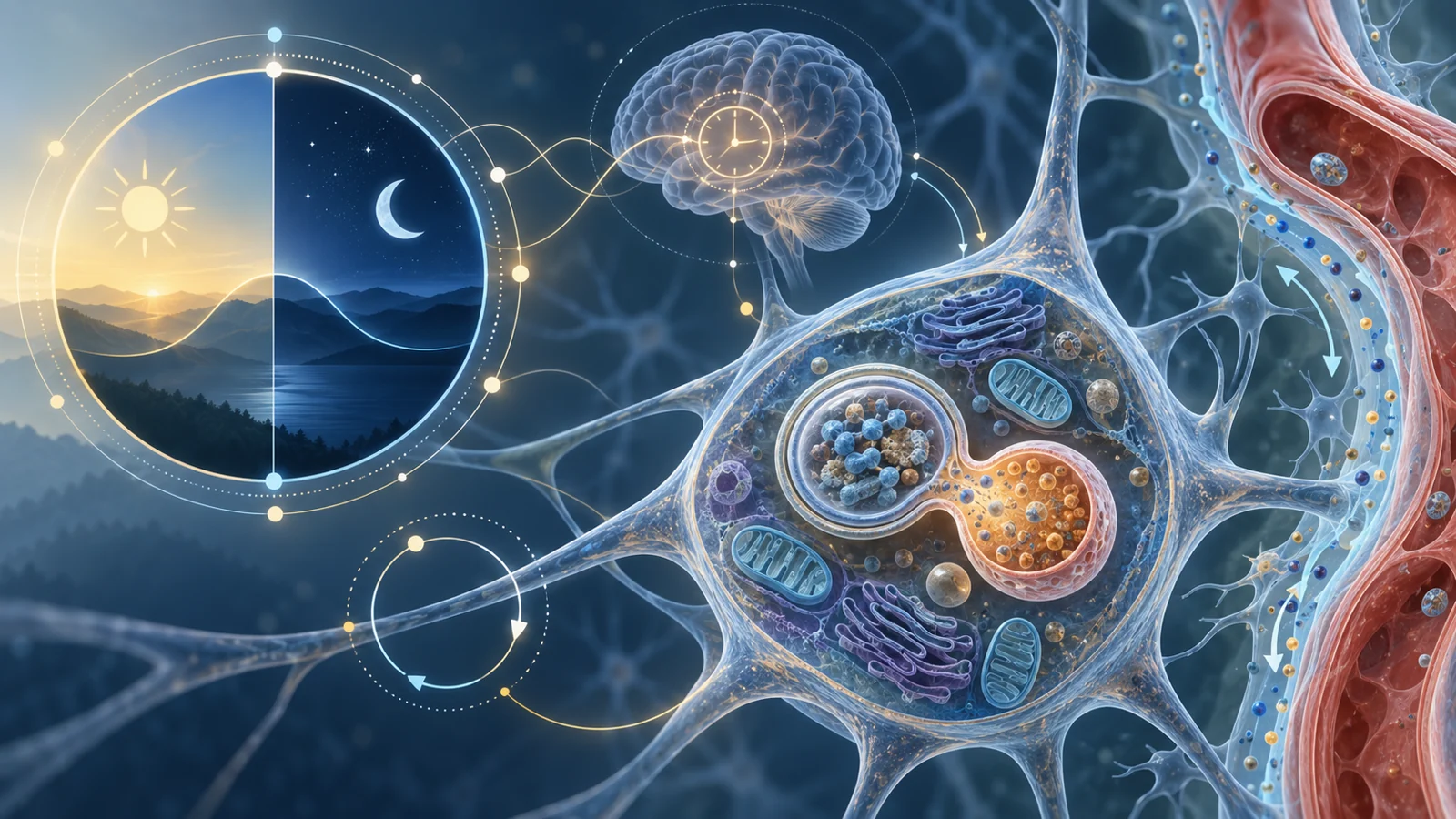
The brain’s central circadian clock sits in the suprachiasmatic nucleus, a small region above the optic chiasm. Light reaching photosensitive cells in the retina provides an important timing input. The central clock then helps coordinate daily patterns that include melatonin and cortisol release, body temperature, and responses to cellular stress.
That system is not simply a record of how many hours someone slept. It is a feedback system that changes gene activity over time. The review describes CLOCK and BMAL1 proteins working together to activate the transcription of other clock genes, including Period and Cryptochrome genes. PER and CRY proteins then form complexes that feed back to restrain CLOCK:BMAL1-driven transcription. Other regulators, including REV-ERBα and RORα, help control rhythmic BMAL1 expression.
The result is a molecular oscillation. Light can reset the system from outside the cell, but the timing pattern is carried through interacting proteins and changes in gene activity. That gives clock-gene function a different biological meaning from sleep duration alone. A person may experience fragmented sleep, while cells may also be receiving altered timing signals that affect when maintenance processes are activated.
Observation and intervention tell different stories
The review contrasts two kinds of experimental evidence. In Alzheimer’s disease mouse models, researchers observed changes in the circadian expression of clock-gene RNA. Altered BMAL1 oscillation has also been reported in people with Alzheimer’s disease. These findings suggest that the clock system changes in the setting of neurodegeneration, but they do not by themselves show whether the changes contribute to disease, result from it, or reflect a feedback process between the two.
The fruit-fly experiments asked a more intervention-like question. Several arrhythmic mutants showed shorter lifespan or faster age-related movement problems, with the ClkAR mutant showing particularly pronounced locomotor deficits. When Clk function was restored, the movement deficits were rescued in the flies.
That rescue is important because it involves changing clock-gene function rather than merely measuring a difference. It suggests that clock activity can influence an observable neurological outcome in an animal. The outcome was movement in Drosophila, however, not human memory or Alzheimer’s disease. Altered RNA in a mouse model and rescued movement in a fly therefore answer different questions. One identifies a changed biological state; the other tests whether altering one component can change a measured phenotype.
The review also summarizes findings in which disrupted circadian timing and sleep fragmentation appear alongside oxidative stress, mitochondrial injury, and impaired waste removal. These relationships help explain why sleep-associated dementia research may point inward toward cellular maintenance, but they do not create one proven causal chain for every form of cognitive decline.
Sleep moves waste outside cells
One pathway discussed in the review involves the glymphatic system, a fluid transport route that follows a network around brain blood vessels. During sleep, this perivascular flow is involved in clearing metabolic waste. Sleep loss can interfere with that process, and the review connects impaired sleep with reduced removal of substances such as amyloid-beta, tau, and alpha-synuclein in the context of neurodegenerative disease.
This is a movement-of-fluid problem around cells. It should not be confused with autophagy, which is a recycling process inside cells.
In macroautophagy, material from the cell, including damaged or unwanted components, is enclosed in a membrane structure called an autophagosome. The autophagosome then joins with a lysosome, an organelle containing the machinery needed to break down and recycle that material. Other forms of autophagy use different routes, but the shared purpose is intracellular housekeeping.
The review describes sleep loss as affecting autophagy proteins and memory formation in the hippocampus. It also summarizes animal-model findings in which circadian control of autophagy was needed to reduce amyloid accumulation and memory impairment. Those findings make the inside-the-cell connection more concrete: daily timing may influence when recycling is available to help maintain neurons.
Still, glymphatic transport and autophagy are not two names for the same mechanism. One concerns fluid movement through a brain-wide perivascular network. The other concerns the sorting and degradation of material within individual cells. Sleep disruption could affect both, but the cited examples do not establish that one universal sequence links sleep loss, failed recycling, amyloid buildup, and dementia in all settings.
Recycling depends on more than one switch
The review’s discussion of mTOR and SIRT1 shows why a clock-linked treatment would be difficult to design. Autophagy is not an isolated lever that can simply be pushed upward. Its activity is connected to broader systems governing growth, stress responses, energy use, and cell survival.
mTOR is presented as a central component of the pathway connecting circadian regulation with autophagy. SIRT1 is also linked to autophagy and has an inverse relationship with mTOR in some of the examples discussed. Under nutrient-limiting conditions, SIRT1 activity accompanied by mTOR inhibition was associated with neurite outgrowth and greater neuronal survival. That example supports the idea that reducing one pathway’s activity can help cells in a particular context.
Other settings are less simple. In circumstances involving dopaminergic neuronal loss, the review describes a need for a balance among SIRT1, mTOR, and FoxO activity to achieve neuroprotection. FoxO proteins can also influence SIRT1 transcription, creating feedback rather than a one-way chain. SIRT1 and FoxO3a have been described as working together in contexts involving amyloid injury, mitochondrial dysfunction, and oxidative stress.
These comparisons matter because a pathway that supports recycling in one biological state may interact differently with growth signals, nutrient conditions, or neuronal stress in another. Increasing autophagy, inhibiting mTOR, or changing a clock gene cannot be treated as universally beneficial without knowing which cells are affected, when the intervention acts, and how the surrounding regulatory network responds. The review presents biological balance as a central therapeutic problem, not a minor technical detail.
The hard step is reaching human cognition
The experiments summarized in the review span several levels of evidence. Clock-gene RNA measurements ask whether molecular timing has changed. Fly movement asks whether restoring clock function can alter an animal phenotype. Neuronal survival and autophagy findings ask how cells respond to stress or altered regulation. Amyloid accumulation and memory performance in animal models move closer to disease-relevant outcomes, but they remain animal outcomes.
A dementia treatment would have to answer a different question: whether a defined intervention can preserve or improve cognition in people while remaining safe over sustained use. That requires more than demonstrating that a gene pathway is involved. It requires an intervention, a treatment exposure, a measurable clinical outcome, and evidence that any benefit is durable enough to matter.
The supplied findings do not establish a patient dosing regimen, treatment effect size, long-term cognitive benefit, or safety profile for clock-linked interventions. They also do not show that correcting sleep disruption prevents dementia or reverses established cognitive loss. Their value is earlier in the chain. They help identify a biological relationship that may be worth testing, while keeping the endpoints attached to the species and experiment that produced them.
The most careful reading is therefore neither that sleep disruption is irrelevant to brain disease nor that clock genes already offer a dementia therapy. The review supports a mechanistic proposal: daily timing may influence several maintenance systems that become important when neurons are under stress. Whether those systems can be targeted without disturbing other essential functions remains an open clinical problem.
What this could change
For people facing cognitive decline, the prospective benefit would be treatments that help preserve brain function by supporting neuronal maintenance. Clock-linked interventions might eventually help cells coordinate recycling, waste handling, and survival more effectively, potentially slowing deterioration. That possibility depends on finding ways to protect the relevant cells and then demonstrating safe, sustained cognitive benefit in people.
The 2021 review points toward that future by connecting daily timing with intracellular housekeeping and neuronal resilience. The experiments summarized in the review suggest where a treatment might act, but do not establish a therapy. If the relationships among clock genes, autophagy, mTOR, SIRT1, and FoxO pathways can be understood well enough to tune them safely, research could move from describing disrupted rhythms toward preserving brain function during cognitive decline.
Key Sources
- Primary source: Cognitive Impairment and Dementia: Gaining Insight through Circadian Clock Gene Pathways.
DOI or Journal Page · Review or Meta-Analysis · 2021-07-09
Keep exploring