
Research Signal – Source date 2026-04-08
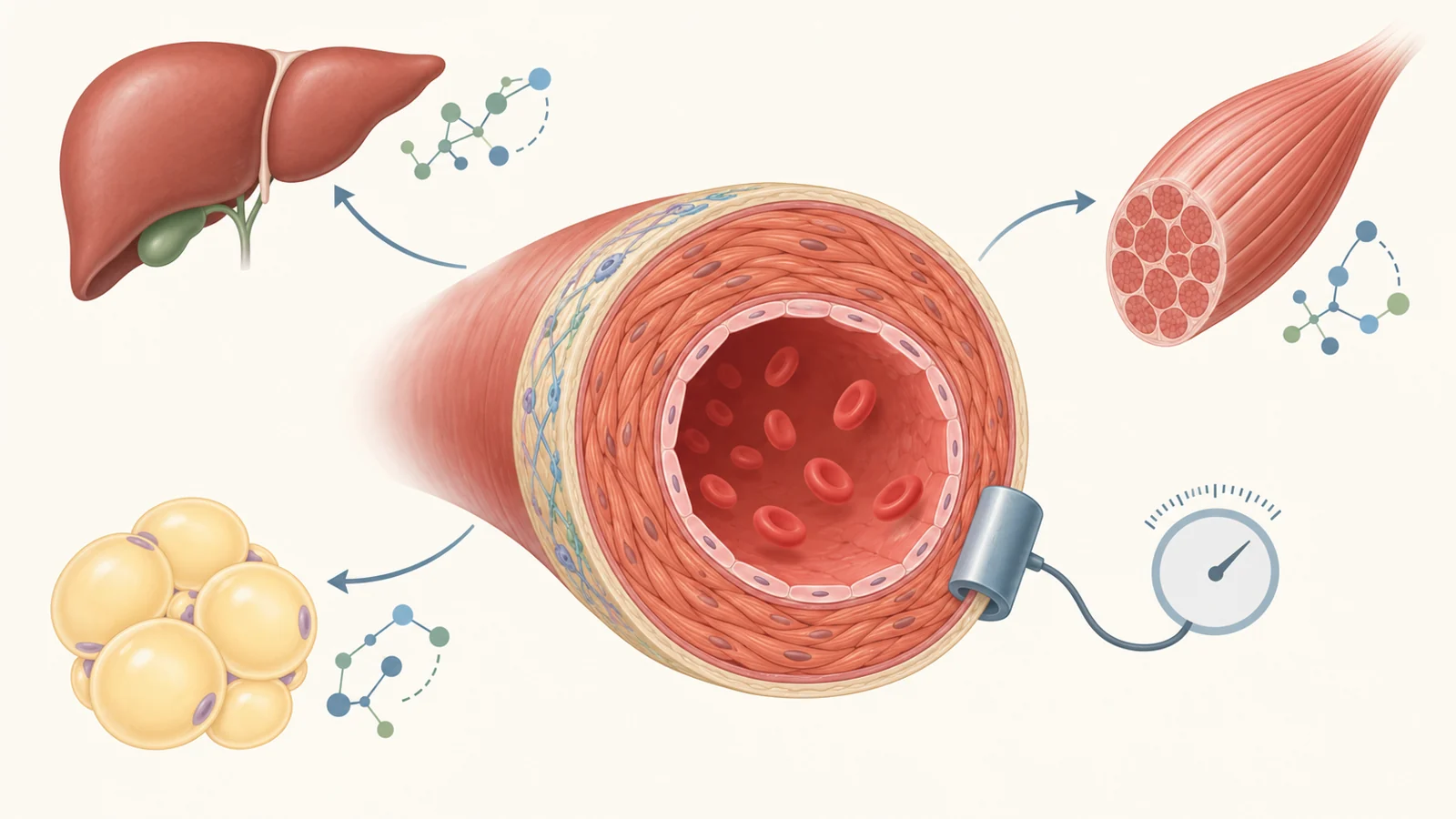
Blood vessels change with age. Their walls can become stiffer, the inner and middle layers can thicken, and the open channel can widen. Cells that help maintain the wall may also decline or stop working well. These changes raise cardiovascular risk, but they are not simply a one-way consequence of time: the 2026 review describes glucose and fat metabolism as both influencing vascular aging and being altered by it.
That two-way relationship matters because a metabolic problem may affect the vessel wall while changes in blood vessels may also affect how organs handle sugar and fats. The review brings together findings from cells, animals, observational research and intervention studies. Those findings help explain possible connections and point to measures clinicians can assess, but they do not show that changing one metabolic pathway reverses vascular aging in people.
Energy use shapes the vessel lining
The endothelium is the thin layer of cells lining the inside of blood vessels. It helps regulate the vessel’s interaction with blood and supports repair. In these cells, glycolysis—a way of breaking down glucose to produce energy—supplies a large share of ATP, the cell’s usable energy. The review says reduced glycolysis can limit endothelial-cell growth and movement, while growth signals such as VEGF and FGF-2 can increase glycolytic activity.
This energy balance is connected to the lining’s ability to maintain blood-flow responses. The review describes a pathway in which glycolysis supports signaling from blood-flow-related purinergic receptors to eNOS, an enzyme involved in producing nitric oxide. It also notes that activation of the P2Y1 receptor promotes endothelial autophagy, a cell-maintenance process that declines with age.
Fat metabolism matters to these cells too. The enzyme CPT1 helps cells use fatty acids; in experimental work summarized by the review, its deficiency reduced endothelial-cell growth and increased permeability. Fat metabolism also affects acetyl-CoA, a molecule used in cellular processes. In cultured human endothelial cells, inhibiting ATP citrate lyase reduced acetyl-CoA production and accelerated senescence, while adding acetyl-CoA delayed senescence induced by hydrogen peroxide. These are cell findings, not evidence that supplementation prevents artery aging in people.
The review also describes how elevated homocysteine can disrupt endothelial cells. At sub-lethal levels, it impaired cell growth by inhibiting mitochondrial respiration, with glycolysis increasing in compensation to help sustain energy. More severe elevation was linked to endothelial-cell death. These examples show why a single label such as “high blood sugar” or “high blood fats” cannot capture every metabolic route affecting the vessel lining.
Smooth muscle can change its role
Smooth-muscle cells make up much of the vessel wall and help it perform its physical functions. They are adaptable: in vascular disease and aging, they can shift their characteristics rather than remaining in one stable state.
The review describes evidence from human narrowed carotid arteries and wire-injured mouse arteries in which HIF-1α and its target PFKFB3 were associated with this switching. The pathway links glycolysis to FASN-mediated fat metabolism. It offers a specific example of glucose and lipid processes acting together as smooth-muscle cells change their behavior in a diseased or injured vessel.
Such cell-level findings help explain how metabolism could shape the wall, but they do not by themselves show how a treatment will affect a person’s arteries. The broader point is that the vessel’s response depends on several cell types and processes, not one metabolic switch.
The loop reaches beyond the arteries
Blood vessels also influence the tissues they supply. The review describes age-related changes in liver endothelial cells, including impaired transfer of insulin across the liver’s specialized vessel lining. Studies in older rats found less insulin reaching liver tissue, alongside reduced hepatic insulin and glucose uptake and weaker activation of insulin signaling. These findings were linked to poorer systemic insulin sensitivity and glucose handling.
The liver examples also connect vessel structure with blood fats. In older adults, clearance of chylomicron remnants—particles left after fat transport—was impaired, accompanied by higher triglycerides after meals. Work in perfused rat livers found that transfer of lipoproteins across liver endothelial cells nearly stopped with age. This suggests a route by which aging vascular changes may contribute to altered lipid levels, though the experimental observations do not establish a single cause of age-related dyslipidemia in people.
Skeletal muscle offers another link. In a mouse obesity model, endothelial p53 was elevated. Genetically reducing p53 in those cells improved insulin resistance and increased muscle glucose uptake through higher GLUT1 expression. The review connects this result with eNOS and a pathway involved in mitochondrial biogenesis. It is an animal finding, but it illustrates how vascular-cell behavior may affect metabolism in a distant tissue.
Measurements capture different parts of aging
Clinicians and researchers can assess vascular aging through measures of structure and function. Pulse wave velocity (PWV) estimates how quickly a pressure pulse travels through arteries and is used to assess arterial stiffness. Carotid-femoral PWV is the predominant approach discussed in the review; brachial-ankle PWV and the cardio-ankle vascular index are alternatives. Carotid intima-media thickness measures the thickness of the carotid artery wall, while flow-mediated dilation assesses how the brachial artery responds after a temporary occlusion. These measures answer different questions about stiffness, structure and endothelial function.
Molecular markers are a different kind of evidence. The review discusses cellular and metabolic processes associated with vascular aging, but it emphasizes that the sensitivity and specificity of proposed diagnostic approaches remain to be established. A stiffness measurement does not identify the cellular pathway behind it, and a proposed molecular clue is not automatically a validated diagnostic test. The supplied evidence does not establish one biomarker that reliably diagnoses vascular aging across populations.
That distinction matters for interpreting treatment studies: a change in one measure may be useful, but it cannot stand in for every aspect of vascular health or for a demonstrated reduction in clinical events.
Interventions show measured improvements, not rejuvenation
Exercise is associated with healthier aging in the evidence summarized by the review. In aged mice, exercise reduced senescence and components of the senescence-associated secretory phenotype, a collection of inflammatory signals, in serum and heart. It also mitigated diet-related senescence in mouse fat and liver. In people, regular physical activity was associated with lower markers of endothelial and immune-cell senescence. These findings span animal experiments and human associations, with different outcomes measured in each.
The review also cites glucose-lowering treatment studies. Metformin, commonly used for type 2 diabetes, was associated in the REMOVAL trial with a slower increase in carotid intima-media thickness: 0.012 millimeters per year less progression over five years among participants with diabetes. The review describes SGLT2 inhibitors as improving vascular phenotypes, and reports that empagliflozin reduced carotid-femoral PWV in people with type 1 diabetes. Combined insulin and metformin treatment was associated with greater improvement in endothelial function than metformin alone in people with type 2 diabetes. These are specific vascular measures; they do not demonstrate that treatment reversed vascular aging as a whole.
For lipid-lowering treatment, the review describes statins as improving endothelial function and potentially slowing arterial stiffness progression in some settings. It also contrasts studies with different treatment intensity and outcomes: low-dose simvastatin lowered LDL cholesterol by 25% without changing carotid intima-media thickness, while high-dose atorvastatin lowered LDL cholesterol by 45% and reversed the reported progression of that thickness measure. The review suggests structural changes may require more intensive and prolonged treatment, but a shift in one artery measure is not the same as reversing the aging process.
The evidence therefore ranges from experimental mechanisms to associations and treatment-related changes in intermediate measures. It does not test one unified strategy that selects people by combined metabolic and vascular markers and then demonstrates better clinical outcomes. Multi-omics research and validation across populations would be needed to establish whether such measures can reliably identify distinct patterns of risk.
How metabolic clues could sharpen vascular prevention
Reliable combined measures of metabolism and vascular health could help clinicians identify people at risk earlier and tailor established prevention choices to the pattern of disease they have. That could make prevention more precise for older adults whose vascular stiffness, glucose handling and lipid patterns do not follow the same course. The benefit remains conditional: these measures must predict risk across populations, and studies must show that tailoring care with them improves outcomes. For now, the review offers a connected account of plausible pathways and measured vascular changes, not proof that a targeted metabolic strategy rejuvenates arteries.
Key Sources
- Primary source: Glucose and Lipid Metabolic Mechanisms in Vascular Aging and Related Therapeutic Strategies.
DOI or Journal Page · Review or Meta-Analysis · 2026-04-08
Keep exploring