
Evidence date · 2025-03-03 · Research note 3 of 3
1 2 3
See current Track status →
Since the previous research note · Partly supported
Track continuity
Since the previous research note: What 80 reported cases reveal about confusing inflammation with cancer progression
Previous expectation: Better ways to distinguish sarcoid inflammation from cancer progression could help teams make treatment decisions.
What this study adds: A 2025 case report documents skin biopsies showing granulomas, a separate pelvic mass showing metastatic cancer, and continued avelumab after a surgery-related pause. Immune-cell mapping adds a tissue comparison but does not validate a general diagnostic method.
What changed in this study
A woman receiving avelumab, a cancer immunotherapy drug, developed two small skin bumps that turned out to be granuloma-forming inflammation. Around the same time, a separately removed pelvic mass was confirmed as metastatic colon cancer. The different diagnoses mattered because either kind of lesion can raise concern about cancer progression, yet the skin biopsies and the pelvic surgery answered different questions.
A 2025 case report described the patient, a 56-year-old woman with metastatic colon cancer, and compared immune cells in her skin lesion and pelvic tumor. The comparison revealed some spatial similarities as well as clear differences. It offered a close look at one unusual reaction during treatment, but it did not establish a general test for telling inflammation from cancer.
Two skin biopsies showed granulomas
The patient entered a phase I trial of avelumab in 2017 after her cancer had progressed through several treatments, including other immunotherapies. Avelumab blocks PD-L1, a checkpoint protein that helps restrain immune activity. By releasing some of that restraint, checkpoint drugs can help immune cells respond to tumors, but immune activation can also accompany inflammation in healthy tissue. The authors interpreted her skin reaction as related to avelumab; the case could not prove exactly how the drug produced it.
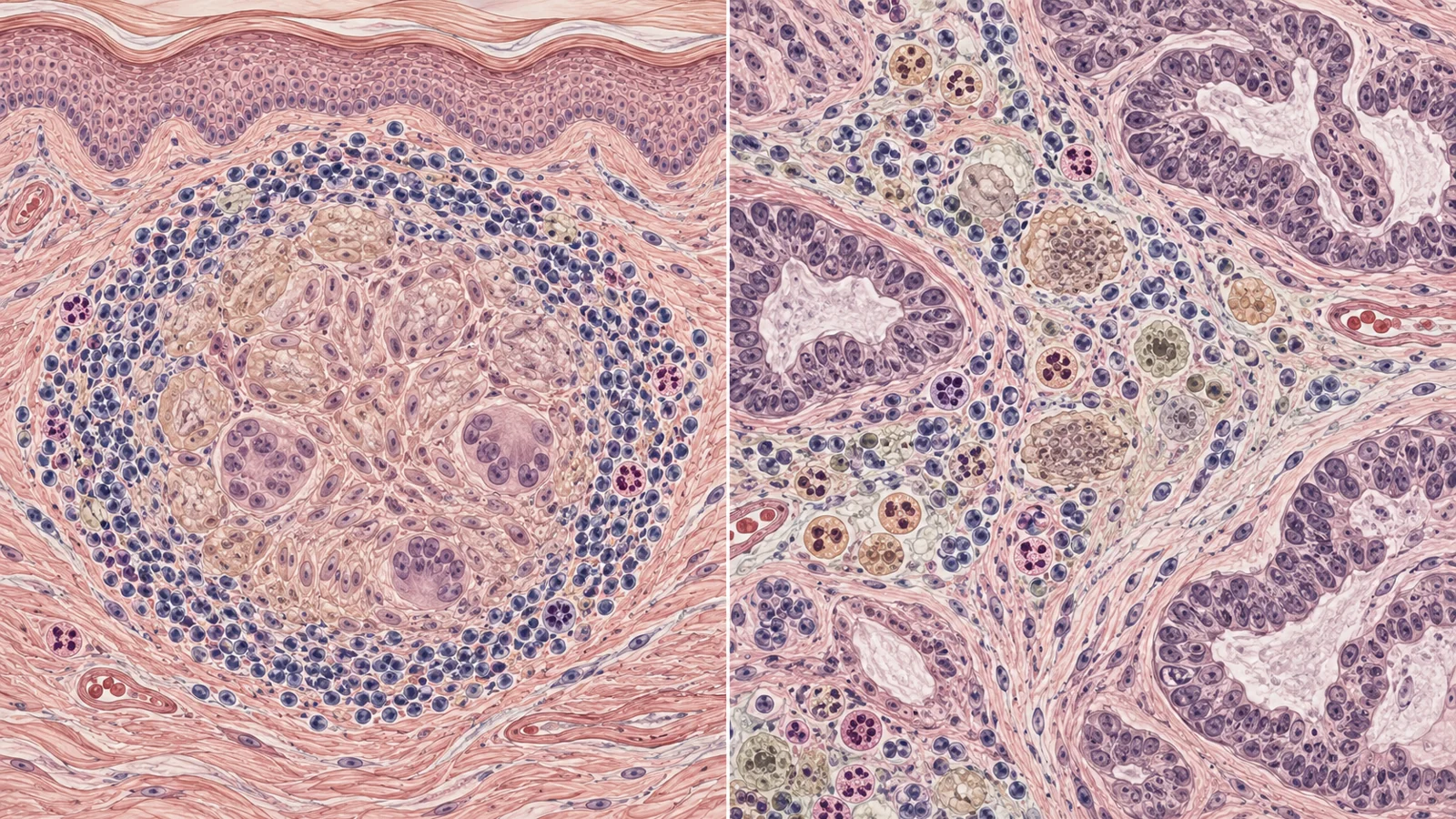
After 30 doses, the patient noticed a small bump on her chin. It had appeared about two months earlier, grown, and sometimes itched. A shave biopsy found non-necrotizing granulomatous inflammation. Granulomas are organized clusters of immune cells that form during some inflammatory reactions. The biopsy was consistent with a sarcoidosis-like reaction, a pattern of granuloma-forming inflammation.
Treatment was then suspended while surgeons removed a pelvic mass. Its pathology showed metastatic adenocarcinoma, meaning cancer had spread from the colon, and found no granulomas in that mass. After recovering from surgery, the patient resumed avelumab. Five doses later, a second skin bump appeared on her upper arm. A biopsy also showed well-formed granulomas without necrosis, with multinucleated giant cells containing asteroid-shaped structures. Taken together with the chin biopsy and treatment timing, the authors diagnosed cutaneous sarcoidosis associated with immunotherapy.
The pathology therefore distinguished the two sampled sites: the skin lesions showed granulomatous inflammation, while the pelvic mass was metastatic cancer. It did not identify the cause of every other abnormal area seen on the patient’s scans.
The immune cells shared a pattern at the edges
To compare the tissues, the researchers used multiplex immunofluorescence, a method that stains several cell markers in different colors so researchers can see which immune cells are present and where they sit. In the skin, CD68-positive macrophages—immune cells that gather in the granulomas—formed tight clusters. Around some granulomas were CD4-positive T cells, CD8-positive T cells, natural killer cells, and regulatory T cells.
The pelvic tumor had no sarcoidal granulomas. Its macrophages formed looser nests around tumor tissue, and were less tightly packed into the rounded, giant-cell structures seen in the skin. Other immune cells surrounded and entered those nests. Even with that different organization, the researchers saw parallels in how immune cells were arranged near the edges of the skin granulomas and around the tumor.
Cell counts also differed between the samples. The skin granulomas had higher densities of CD68-positive macrophages and more M2 macrophages, both within the granulomas and in nearby tissue. They also contained more CD8-positive T cells than the metastasis. In tissue around the skin granulomas, natural killer cells were more numerous than in tissue around the pelvic tumor. The pattern is a comparison within this patient, not evidence that these cell arrangements identify sarcoidosis or cancer in other people. It also does not explain what triggered the reaction.
The chest nodes remained a separate question
The patient also had enlarged lymph nodes in her chest, an area where sarcoidosis-like reactions can occur. But these nodes were present before she started avelumab and had never been biopsied. The authors considered their timing and course more consistent with her metastatic cancer, while acknowledging that their tissue identity could not be confirmed.
Imaging alone could not settle the question. Both sarcoidosis and cancer can show increased uptake on PET scans, so a scan’s bright signal is not a tissue diagnosis. The skin biopsies established what was happening in the sampled skin; they could not show whether the unbiopsied chest nodes contained inflammation, cancer, or both.
Surgery and skin treatment followed different timelines
The pause in avelumab followed the need for pelvic surgery. After recovery, treatment resumed, and the arm lesion appeared after five additional doses. For the skin reaction, the patient received topical fluocinonide ointment once daily because the persistent chin lesion’s appearance distressed her. Four months later, both skin lesions were stable.
The report describes the cancer as stable during the two-year, 56-dose avelumab treatment protocol. That course occurred in one patient, whose care also included surgery and earlier treatments. It cannot establish which approach would be best for someone else, or whether a sarcoidosis-like reaction predicts a cancer outcome.
Why telling inflammation from cancer spread could protect treatment choices
More reliable ways to recognize sarcoid-like reactions could help oncology teams judge whether a new lesion signals cancer progression before changing a treatment that may still be useful. This case shows why the distinction can matter: the skin biopsies and pelvic surgery found different conditions in the same patient, while the chest nodes remained unresolved. Turning that insight into a dependable diagnostic approach and clearer treatment decisions will require evidence beyond a single case.
Key Sources
- Primary source: Case Report: The immune architecture of immunotherapy-induced cutaneous sarcoidosis resembles peritumoral inflammation.
DOI or Journal Page · Clinical Trial · 2025-03-03