
Research Signal – Source date 2026-02-01
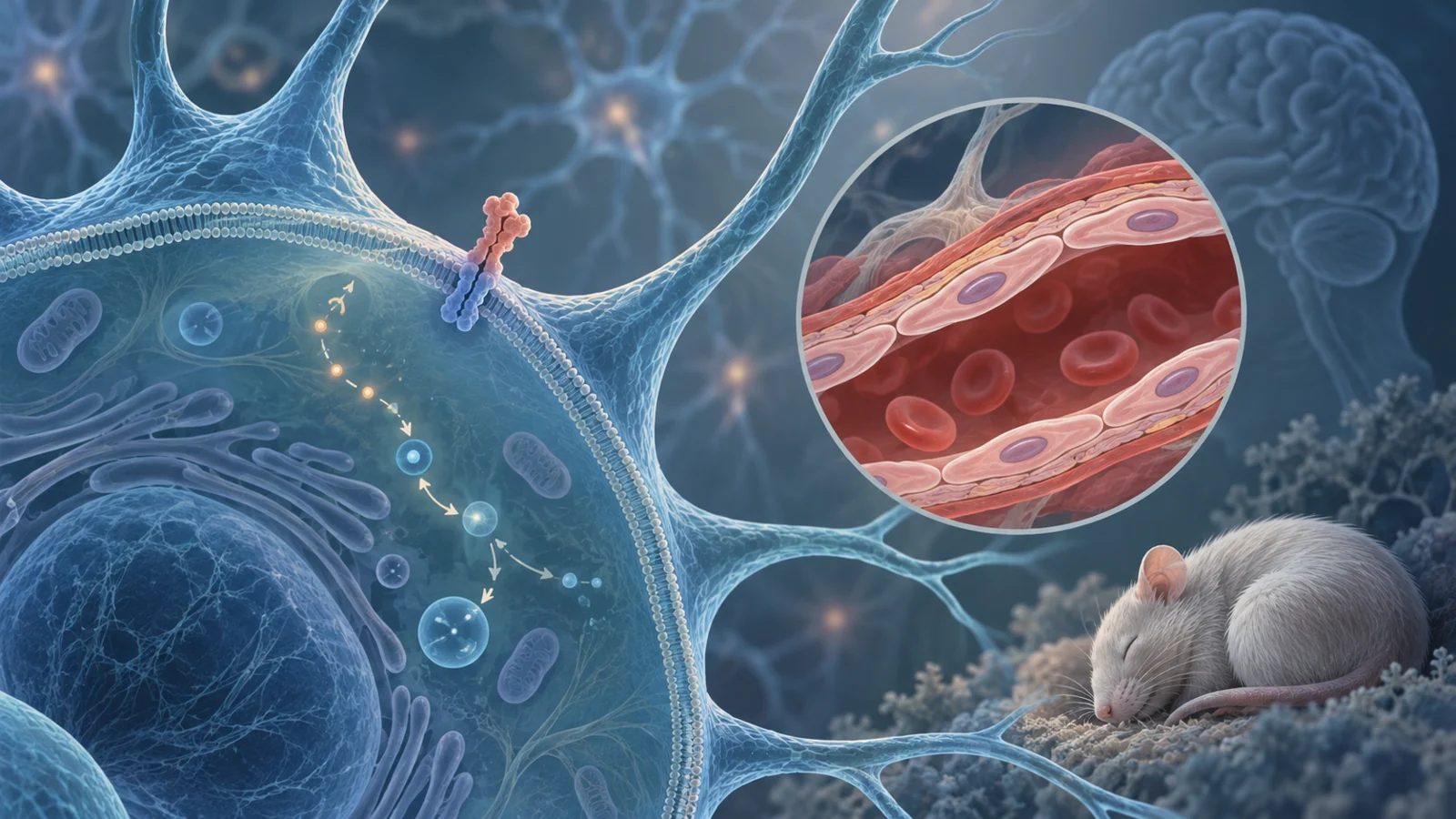
Amyloid precursor protein (APP) is known as the source of amyloid beta, the peptide that accumulates in Alzheimer’s disease. But APP fragments can also participate in signaling inside cells. A mouse study published February 1, 2026, tested whether strengthening one such signal could affect sleep and the blood-brain barrier, the interface that helps regulate what passes between blood and brain tissue.
The experiment’s central result was striking: mice with an Alzheimer’s-like disease model that received a signaling-capable APP fragment had sleep patterns and several other measured outcomes closer to those of non-transgenic mice. A version of the fragment altered so it could not interact with GαS, a signaling protein, did not produce the same pattern. The findings point to a possible protective role for APP signaling, while leaving open how the measured changes relate to one another and whether the approach could matter beyond this mouse model.
APP has another role beyond producing amyloid beta
APP can be cut along different routes. In one, enzymes generate amyloid beta. In another, cleavage prevents amyloid beta formation. The resulting membrane-bound APP fragments include an intracellular portion called AICD, which can participate in signaling. Earlier work by the research team found that keeping an APP fragment at the cell membrane engaged GαS and reduced amyloid pathology in mice. Amyloid burden was not a new outcome measured in this follow-up experiment.
For the new test, the researchers engineered a membrane-tethered APP intracellular domain, or mAICD, designed to keep signaling active. They compared it with a mutant version that lacked the GαS interaction site and with a membrane-targeted control construct. The mutant matters because it probes whether the signaling connection, rather than simply introducing a fragment, is needed for an effect.
The team delivered the constructs by viral injection into the brain ventricles of newborn 5XFAD mice, which carry five familial Alzheimer’s-associated mutations, and non-transgenic littermates. The researchers assessed the animals months later, at about 6 to 8 months of age. This timing means the experiment tested an intervention present from early life, not one started after Alzheimer’s-like changes were already established.
Sleep changed during the dark phase
A sensor beneath each cage detected pressure changes as the mice moved or rested. Software classified short segments of those signals as sleep or wake, providing a continuous, cage-based estimate rather than a direct recording of brain electrical activity. The researchers monitored the animals for 48 hours.
During the light phase, the study found no differences in sleep duration or pattern. In the dark phase, when mice are normally more active, 5XFAD mice slept less than non-transgenic littermates: about 21.9% of the period compared with 31.2%. The 5XFAD mice expressing mAICD showed a significant improvement, with sleep behavior described as remarkably similar to that of non-transgenic mice. The GαS-binding-deficient mutant did not show the same rescue.
This comparison supports a role for the mAICD–GαS connection in the observed sleep effect. It does not by itself explain whether the signal acted on sleep-regulating circuits directly or through other changes in the brain.
A preference test offered one view of memory
To assess recognition memory, mice first encountered two identical objects and later saw one familiar object alongside a new one. The researchers calculated a preference score from the time spent near the novel object relative to time near both objects. It is a specific behavioral measure: it reflects the animals’ tendency to explore something new, not memory in every sense.
On this test, 5XFAD control mice had a lower novel-object preference score than non-transgenic controls, 46.4% versus 68.9%. The score in 5XFAD mice expressing mAICD was 74.6%, while the mutant group’s score was 54.6% and did not show the same improvement. These group comparisons are distinct from the study’s correlations across individual mice. Across the animals, better sleep scores were associated with higher novel-object preference, but that relationship cannot show that improved sleep caused better performance.
The barrier result came from one injected tracer
To estimate blood-brain barrier permeability, the researchers injected fluorescent dextran into the mice’s bloodstream, then measured how much accumulated in brain tissue. The molecule used was 3 to 5 kilodaltons in size. More tracer in the brain indicated greater passage of that particular probe under the assay conditions.
Compared with non-transgenic controls, 5XFAD control mice had 1.45 times as much brain tracer. The mAICD group had a lower level, 1.06 times the control reference, while the mutant group remained elevated at 1.56 times. The result is evidence about permeability to this dextran tracer; it does not establish restoration of every barrier function or component.
Across individual mice, tracer permeability was inversely correlated with sleep and with novel-object preference. These associations show that the measures varied together in this experiment, but they do not establish which change came first or whether one caused another. Sleep, barrier permeability and behavior could be linked through the APP signal or through additional processes.
Astrocytes appeared differently positioned near vessels
The researchers also examined GFAP, a marker used to visualize astrocytes, cells that help support the brain’s neurovascular units, including its small vessels. In 5XFAD mice expressing mAICD, GFAP staining was lower in the cortex and hippocampus than in 5XFAD controls. The investigators also saw differences in astrocyte shape and distribution, including more GFAP-positive area associated with neurovascular units in mAICD-expressing animals.
These images offer a possible lead about how the local cellular environment around vessels may change alongside the tracer result. They do not identify which cell type produced the relevant signal or show that astrocyte redistribution caused the barrier measurement to improve. The viral construct could reach multiple cell types, and the study did not isolate their individual contributions.
Some patterns varied between male and female mice
The researchers analyzed males and females separately and found that certain sleep and behavioral relationships differed by sex. For example, the association between sleep and novel-object preference was evident in females but not males; the link between barrier permeability and open-arm behavior appeared stronger in males. The reduction in tracer permeability associated with mAICD was comparable across sexes.
These results caution against treating the group averages as a single uniform response. They also leave open how sex, hormonal variation and genetic background may have shaped the measurements in this mouse cohort.
A possible path to protecting sleep and memory in Alzheimer’s
If APP-mediated signaling can be made to protect sleep and vessel-barrier function when activated after disease changes begin, it could eventually point toward ways to preserve brain function in Alzheimer’s disease. That possibility depends on major unanswered questions: the effect here followed neonatal gene delivery in one mouse model, and the experiment’s tracer assay captured only one aspect of barrier permeability. The observed correlations also do not resolve whether sleep, barrier function or memory is the first part of the system to change. For now, the study offers a biological lead, not evidence of a treatment for people.
Key Sources
- Primary source: APP-mediated intracellular signaling rescues sleep impairment and blood-brain barrier leakage in Alzheimer's disease mouse model.
DOI or Journal Page · Peer-Reviewed Paper · 2026-02-01
Keep exploring