
Research Signal – Source date 2021-08-16
Someone with Alzheimer’s disease may sleep poorly at night and still struggle to stay awake during the day. Those symptoms can seem contradictory, but they can arise when the brain systems that coordinate sleep and alertness are damaged in different ways.
A review published on August 16, 2021, examines how this might happen. It brings together findings from animal studies of sleep and wake circuits, human postmortem examinations, and observations of sleep. The authors argue that tau, a protein that can form abnormal tangles inside neurons, may affect sleep-regulating regions early in Alzheimer’s disease. The proposal helps connect sleep changes with the underlying disease, but it does not make every sleep problem a specific sign of Alzheimer’s or establish tau as the only cause.
One disease can unsettle both sides of the daily cycle
Sleep disturbances are reported in around half of people with Alzheimer’s disease in the literature reviewed. Changes can appear during preclinical stages, sometimes one or two decades before noticeable cognitive decline, and often become more severe as the disease progresses. That timing is important to the review’s argument, but it is not a diagnostic timetable for an individual.
The pattern is also broader than simply sleeping less. People with Alzheimer’s may spend less time asleep overall, lose a significant amount of slow-wave sleep, or deep sleep, and experience less REM sleep. Nighttime awakenings become more frequent, reducing sleep efficiency and fragmenting rest. During the day, increased sleepiness can make it difficult to remain alert. In advanced dementia, the rhythm of sleep and wakefulness can also lose strength and shift later.
Some changes in sleep are part of ordinary aging. Older adults generally experience less total sleep, more light sleep, fewer periods of deep and REM sleep, and more shifts between sleep stages. The disturbances described in Alzheimer’s can resemble an intensified version of that pattern, but the disease can also involve more substantial disruption of the systems that organize the cycle. Sleep complaints alone therefore cannot identify Alzheimer’s disease or predict future dementia.
A coordinated network decides when the brain changes state
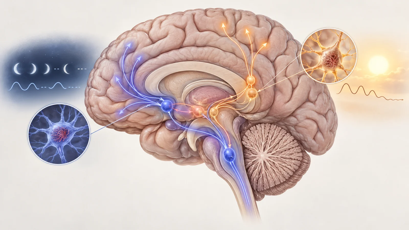
Sleep and wakefulness are not controlled by a single switch in one location. They emerge from an interconnected set of neuronal populations in the brainstem, hypothalamus, and basal forebrain. These regions send broad projections through the brain and influence whether the cortex is organized for alertness, deep sleep, or REM sleep.
The review describes a proposed arrangement sometimes called a flip-flop switch. Wake-promoting and sleep-promoting populations inhibit one another, allowing the brain to settle into one state rather than remain indefinitely between them. Circadian signals from the suprachiasmatic nucleus help set the timing, while the separate sleep and wake populations help determine the state itself.
Wake-promoting neurons are largely excitatory and help maintain arousal. Sleep-promoting populations are often GABAergic, or help activate GABAergic neurons that suppress wake-promoting activity. Damage to wake systems could make alertness unstable and increase daytime sleepiness. Damage to sleep-promoting systems could make sleep difficult to initiate or maintain. If several parts of the network are affected, a person could lose both consolidated nighttime sleep and sustained daytime wakefulness.
This functional picture comes largely from work in animal models and from the known organization of these circuits. It provides a way to interpret the human pathology, but a circuit effect demonstrated experimentally in animals is not by itself proof of the same causal chain in human Alzheimer’s disease.
Tau reaches wake-promoting regions early in the disease sequence
The review’s timing argument focuses first on regions that help keep the brain awake. In the authors’ account, abnormal tau appears in several wake-promoting areas before amyloid-beta plaques become prominent, while sleep complaints may already be present before cognitive symptoms. The overlap in timing leads them to propose that tau-related damage is an important early contributor to disturbed sleep and wakefulness in Alzheimer’s disease.
The locus coeruleus is one example. It is a brainstem center containing noradrenaline-producing neurons that support arousal and other broad brain functions. Human postmortem studies summarized by the review report abnormal tau tangles, loss of neurons, and reductions in the size of the locus coeruleus in Alzheimer’s disease. The amount of tangle pathology, neuronal loss, and volume reduction also tends to track disease progression in the reviewed findings.
Those observations are related, but they are not interchangeable. Finding abnormal tau inside neurons is a different measurement from counting fewer surviving neurons, and neither observation directly measures a person’s sleep on its own. The proposed link to early sleep disruption comes from bringing the timing, anatomy, and clinical pattern together.
The lateral hypothalamic area offers a second example. It contains orexin-producing neurons, which are active during wakefulness and project to several arousal centers, including the locus coeruleus. Experimental loss or inhibition of these neurons destabilizes wakefulness and can produce a narcolepsy-like state in animal work. In Alzheimer’s tissue described by the review, the lateral hypothalamic area shows abnormal tau, loss of total neurons, and loss of orexin-producing neurons. The region is affected at relatively early stages of the pathology described by the authors.
Amyloid remains part of the picture. The review does not argue that tau explains every later sleep disturbance or that amyloid-beta has no role. Instead, it suggests that early tau-related injury to sleep and wake circuits may help initiate the problem, with amyloid and other disease processes contributing through additional pathways as Alzheimer’s progresses.
PSP shows why the affected circuit matters
Progressive supranuclear palsy, or PSP, provides a useful comparison because it is a degenerative disease in which tau pathology occurs without amyloid plaques. People with PSP can have profound difficulty falling and staying asleep, large reductions in deep and REM sleep, and a reduced ability to recover lost sleep through daytime napping. The symptoms point to a severely disrupted sleep system, but the pattern is not identical to Alzheimer’s disease.
The review compares pathology in several wake-promoting regions. In many of those regions, Alzheimer’s disease shows more extensive neuronal loss and related damage than PSP, even though both diseases involve tau. The comparison suggests that the type of tau-related disease, the region affected, and the vulnerability of particular cells may all matter. It does not experimentally isolate tau as the sole cause, nor does it establish a uniform relationship between the number of tangles and the number of neurons lost.
The pedunculopontine region is an important exception. In Alzheimer’s disease, it contains a moderate amount of tangle pathology and fewer glutamatergic neurons. In PSP, the reviewed studies report more pronounced neuronal loss, fewer glutamatergic neurons, and heavier tangle pathology in this region. A single region therefore does not follow the broader comparison across wake-promoting areas.
This helps explain why the diseases cannot be ranked by one simple measure of damage. Tau deposits, surviving cell numbers, and the resulting sleep pattern describe different parts of the process. The greater sleep loss in PSP, despite the broader pattern of more severe wake-region damage in Alzheimer’s, leads the authors to suspect that sleep-promoting regions may have a different pattern of vulnerability in PSP. That possibility remained largely untested in the human studies discussed by the review.
The sleep-promoting side has less human evidence
The reviewed evidence is strongest for wake-promoting regions. Only a small number of sleep-promoting areas had been examined in Alzheimer’s postmortem studies, including the intermediate hypothalamic nucleus, the lateral hypothalamic area, and the substantia nigra. Other sleep-promoting populations identified through animal research or some in-vivo work had not yet been examined sufficiently in Alzheimer’s disease.
One human study discussed in the review examined galanin-expressing neurons in the intermediate hypothalamic nucleus alongside sleep-actigraphy measurements in older adults, including some people with Alzheimer’s disease. People with Alzheimer’s had fewer of these neurons, and people with more fragmented sleep also had fewer galanin-expressing neurons. The finding supports the idea that this region contributes to healthy sleep, but the study did not resolve whether tau accumulation drives the loss of those neurons.
The different kinds of evidence answer different questions. Animal experiments can test what happens when a particular circuit is activated, inhibited, or lost. Human tissue can show where tau pathology and neuronal loss occur after disease has developed. Sleep measurements can show how a person’s rest and alertness are organized. These observations can reinforce one another, but they do not become one unified experiment simply because they point toward the same model.
The distinction matters for treatment claims as well. Animal models suggest that disturbed sleep can increase amyloid-beta levels, and the review discusses evidence that dysfunction in the locus coeruleus may influence amyloid accumulation. Those findings raise the possibility of a feedback loop between circuit damage and other Alzheimer’s pathology. They do not establish that treating sleep disruption in a person will prevent cognitive decline, and they do not show that a sleep symptom identifies the specific pathology causing it.
How sleep circuits could guide earlier Alzheimer’s care
The most practical possibility raised by this work is earlier recognition of disease-related change. If future studies reproducibly connect particular losses in sleep- or wake-promoting neurons with specific patterns of fragmented sleep, daytime sleepiness, or altered daily timing, sleep measurements could become one part of a screening approach aimed at detecting disease before cognitive decline. Any such tool would still need clinical validation to show that it is accurate and useful for patients rather than merely consistent with a biological theory.
The same circuit-level understanding could point toward treatments designed for the systems that sustain sleep or alertness. For someone whose nights are repeatedly broken or whose daytime wakefulness is failing, a therapy that restores or stabilizes the affected network could offer a more targeted benefit than treating sleep as an isolated symptom. In the 2021 perspective of this review, that remains a conditional prospect: the links between tau-related pathology, particular neuronal populations, sleep patterns, and later disease consequences must first be reproduced and tested in clinical settings.
Key Sources
- Primary source: Tau-driven degeneration of sleep- and wake-regulating neurons in Alzheimer's disease.
DOI or Journal Page · Review or Meta-Analysis · 2021-08-16
Keep exploring