
Research Signal – Source date 2026-06-01
A positive test for an Alzheimer’s-related protein identifies a biological feature. Explaining which abilities will become impaired, or how quickly they might change, requires more information. Two people with evidence of Alzheimer’s biology can have different patterns of brain involvement and additional disease processes affecting their symptoms.
A research-focused review published on June 1, 2026, examines this gap through studies using the Alzheimer’s Disease Neuroimaging Initiative, or ADNI. Established in 2004, ADNI combines repeated assessments of cognition and clinical health with brain imaging, genetic information and biological samples. Its shared data let researchers examine how changes in one measurement relate to changes in others over time.
The findings connect differences in what tests detect with differences in where pathology develops and what other conditions accompany it. They help explain why identifying Alzheimer’s biology and understanding a person’s disease course are related but distinct tasks. They do not turn a single positive result into an individual timetable for symptoms or decline.
What the measurements actually capture
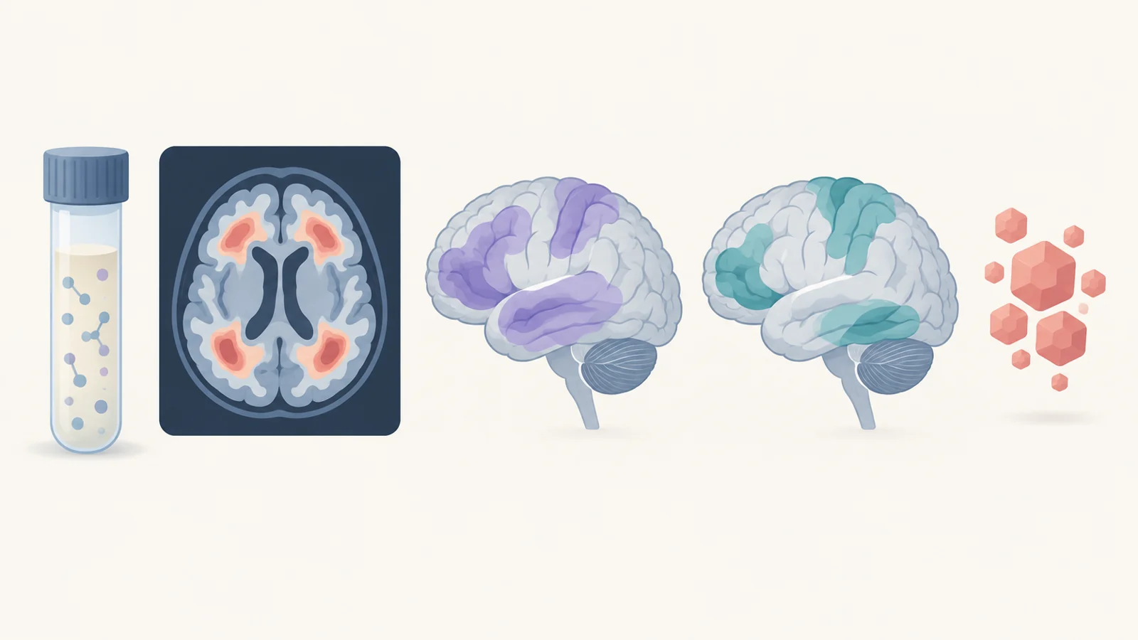
Even tests described as measuring amyloid can be observing different forms of the protein. Amyloid beta is one of the characteristic proteins involved in Alzheimer’s disease. Measurements of amyloid beta 42 in cerebrospinal fluid—the fluid surrounding the brain and spinal cord—reflect its soluble form. Amyloid positron emission tomography, or PET, detects fibrillar aggregates, the protein assembled into deposits.
This distinction gives disagreement between the measurements a biological meaning worth investigating. The fluid result and the scan are not interchangeable readings of exactly the same material. Reviewed studies explored whether the balance between soluble and deposited amyloid relates to genetic factors and cognition. That balance may help researchers understand distinct routes to an abnormal amyloid result.
The same need for precision applies to the broad category of neurodegeneration. MRI measures of tissue loss, measurements of reduced brain metabolism and fluid markers of neuronal injury capture different aspects of deterioration. An anatomical change shows something different from altered energy use or a protein associated with injury to nerve cells.
Combining these measurements therefore offers more than extra chances to detect an abnormality. It lets researchers ask whether changes in protein accumulation, tissue structure and brain function occur together, and where their timing diverges.
Blood vessels and clearance leave a causal puzzle
One possible connection involves the brain’s small blood vessels and its glymphatic system, a fluid transport system implicated in waste clearance. The review describes studies linking small-vessel disease, amyloid burden and cognition with imaging-derived indicators related to this system.
These indicators require careful interpretation. The review reports associations between an imaging-derived measure called the ALPS index and cognition, small-vessel disease and amyloid burden. It is an indirect indicator used to investigate clearance-related function, rather than a direct observation of amyloid being removed from the brain. Other studies used MRI-derived measures of free water to examine related questions.
In one analysis, a free-water measure statistically accounted for part of the relationship between ischemic heart disease and executive function, the abilities involved in planning and managing tasks. Such a result helps identify a possible connection to investigate. Statistical mediation alone cannot establish the biological steps connecting heart disease to cognitive difficulty.
A plausible feedback process could run in both directions. Small-vessel damage might interfere with clearance, while early disease-related changes might themselves disturb fluid transport and the blood–brain barrier, which regulates exchanges between blood and brain tissue. The review leaves these alternatives open.
The vascular findings also resist a simple explanation. Some analyses connected vascular disease with progression. Another found that certain white-matter abnormalities were more closely related to amyloid in vessels and brain tissue, along with neurodegeneration, than to systemic vascular risk after other factors were considered. These differences matter when choosing what a treatment should target. The associations do not establish that improving a clearance indicator or modifying the proposed vascular process would improve Alzheimer’s outcomes.
Where tau accumulates helps explain which abilities change
Tau provides a more anatomical part of the explanation. The review connects tau accumulation in the medial temporal lobe, a region important for memory, with memory impairment. More widespread accumulation was associated with difficulties across additional cognitive abilities and everyday function.
This makes the location of an abnormality consequential. A measurement of overall disease burden can obscure whether pathology is concentrated in a relatively limited region or distributed across areas supporting several abilities.
A study summarized in the review examined abnormal tau deposition across 70 brain regions. More than 80% of participants followed a pattern broadly consistent with conventional tau staging. Yet substantial regional differences remained, especially among participants with mild cognitive impairment.
In that group, tau accumulated particularly quickly, while the affected regions overlapped less between participants than they did among those with Alzheimer’s dementia. The findings suggest that several patterns of cortical involvement may become more similar later in disease progression.
A recognizable staging pattern can therefore coexist with meaningful differences in anatomy during earlier stages. Agreement with that pattern describes the regional organization of tau; it does not establish that everyone follows an identical sequence across all the biological processes involved in Alzheimer’s.
Another pathology can accompany Alzheimer’s changes
Some differences in symptoms and progression may reflect an additional disease process. Lewy body disease involves misfolded alpha-synuclein, a protein distinct from amyloid beta and tau. It can occur alongside Alzheimer’s-related changes.
In the ADNI postmortem cases described by the review, Lewy body pathology was present in around half of individuals with intermediate to high Alzheimer’s neuropathological change. That is a finding about a particular autopsy subgroup, not a prevalence estimate for everyone living with Alzheimer’s.
A cerebrospinal-fluid test called a seed amplification assay now allows researchers to investigate this accompanying pathology in living participants. The review describes its use in detecting misfolded alpha-synuclein in cerebrospinal fluid. It creates a way to compare evidence of that process with symptoms and brain measurements collected during life.
Across the studies summarized, alpha-synuclein positivity was associated with faster cognitive decline, earlier symptom onset and a greater risk of clinical progression. It was also linked to hallucinations and difficulties with executive and visuospatial abilities—the latter involving the interpretation of visual information and spatial relationships.
These observations give accompanying pathology a concrete role in interpreting a person’s presentation. Difficulties involving vision, spatial processing or planning may arise in a biological setting that includes more than the amyloid and tau changes identified by Alzheimer’s tests. Detecting the additional process lets researchers study how it relates to the pace and character of decline.
How much weight the findings can carry
The review draws on a defined selection of research. Its authors searched PubMed, Web of Science and Google Scholar for publications using ADNI data or biospecimens, with an eligible electronic-publication window from January 1, 2023, through June 30, 2025. That window is distinct from the review’s June 1, 2026 publication date.
The search identified approximately 1,830 publications before further exclusions and selection. The research-focused synthesis separated out clinically oriented work and excluded papers devoted primarily to methods or non-Alzheimer’s topics. It also filtered papers by the impact factors of the journals publishing them. The authors acknowledge that this arbitrary cutoff may have left out relevant studies. Journal-level filtering limits which findings enter the account; it does not independently validate each included result.
ADNI’s open-data model is a major strength because different teams can investigate relationships across imaging, samples and clinical assessments. But several analyses may involve overlapping participants. Repeated findings from a shared resource are not automatically independent confirmations in new populations.
Some of the reviewed work included other cohorts, adding opportunities to test whether particular findings travel beyond ADNI. That support has to be assessed study by study. Longitudinal observations can clarify timing, while genetic analyses and statistical models can test proposed relationships, but those approaches do not settle every causal question. The authors also call for validation in more diverse populations, a necessary step before biological groupings developed in research can reliably guide broader clinical use.
What this could change
Reproducible biological profiles could help clinical trials match participants to treatments aimed at the processes contributing to their impairment. A profile combining regional tau involvement, vascular changes and evidence of accompanying pathology could make it possible to test more specific treatment questions than a broad Alzheimer’s label alone permits.
For example, trials could investigate whether participants with a particular contributing process benefit from an intervention directed at it, and whether combining approaches produces meaningful gains. That would connect the measurements to a practical decision about whom a treatment helps.
If such profiles predict treatment response, and the targeted pathways prove modifiable with clinical benefit, patients could eventually receive more precisely matched treatment combinations. The intended payoff would be better preservation of cognition and everyday function. The research reviewed here supplies biological reasons to pursue that possibility; realizing it depends on demonstrating that the distinctions identified in scans and samples lead to better treatment outcomes.
Key Sources
- Primary source: Contributions of the Alzheimer's Disease Neuroimaging Initiative to advancing AD research: a targeted review of recent publications.
DOI or Journal Page · Peer-Reviewed Paper · 2026-06-01
Keep exploring