
Research Signal – Source date 2026-02-01
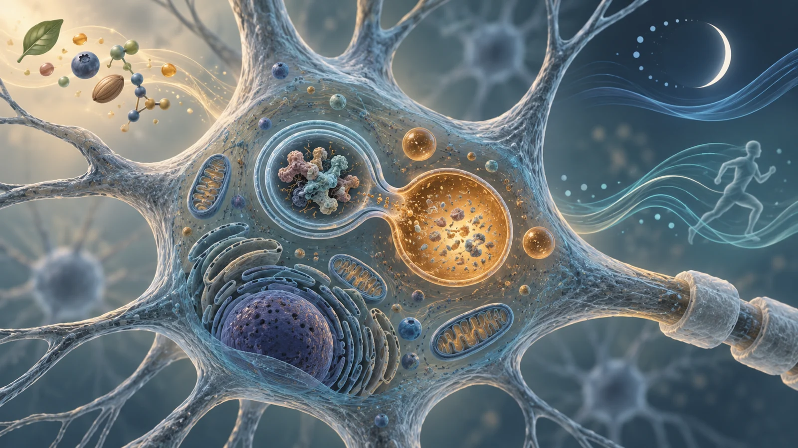
A review published on February 1, 2026, brings together research on three familiar influences on brain health: diet, physical activity, and sleep. Its focus is autophagy, the cell’s system for enclosing, breaking down, and recycling worn-out or damaged material.
That focus matters in Alzheimer’s disease because the disorder is associated with problems in protein homeostasis. Amyloid beta and tau can accumulate, while the cellular systems responsible for handling damaged material may become disrupted. The review asks how everyday physiological states might influence those systems. It also shows why a biologically attractive idea should not be mistaken for a proven prevention strategy.
A recycling system with a throughput problem
Autophagy is not a single switch. In the form emphasized by the review, cellular material is first enclosed inside a double-membrane compartment called an autophagosome. The autophagosome then has to mature and fuse with a lysosome, the cell’s enzyme-filled recycling compartment. Only after that fusion can the cargo be broken down under acidic conditions.
The useful concept is flux: material needs to move through the entire sequence, from collection to delivery to degradation. A cell can form more autophagosomes without successfully completing the process. If fusion with lysosomes fails, vesicles may accumulate while damaged cargo remains incompletely processed. A higher reading of a marker such as LC3 therefore cannot, by itself, show that cellular clearance has improved. It might reflect more autophagosome formation, or a traffic jam later in the pathway.
The same caution applies in the other direction. Autophagy often protects cells under stress, but excessive activation or poorly controlled self-digestion can also damage neurons. The review describes autophagy as part of a wider relationship with oxidative stress and inflammation. Impaired degradation can allow dysfunctional material and reactive oxygen species to build up, while oxidative stress can interfere with the machinery needed for autophagosome–lysosome fusion. In microglia, the brain’s immune cells, autophagy is also connected with the control of inflammatory activity.
The goal is therefore not simply to maximize recycling. It is to restore a workable balance in which damaged material is cleared without pushing cells toward harmful overactivation.
Why eating patterns enter the discussion
Fasting gives researchers a way to study how nutrient availability changes the signals that regulate autophagy. During fasting, cells shift away from relying primarily on incoming glucose and begin using alternative energy sources, including ketone bodies. Nutrient-sensing signals associated with insulin, IGF-1, mTOR, and AMPK change in ways that can favor autophagy and energy maintenance.
Calorie restriction and intermittent fasting are consequently prominent in the review. So are ketogenic, Mediterranean, plant-based, and polyphenol-rich dietary patterns, which may influence overlapping nutrient-sensing and cellular maintenance pathways. These categories are not interchangeable, and the review does not establish a single diet, fasting schedule, or target level of autophagy for protecting the human brain.
The human evidence is also more varied than a simple story about fasting turning on neuronal recycling. One study discussed in the review found increased expression of autophagy-related genes in the skeletal muscle of healthy adults who had practiced long-term calorie restriction. That result is evidence of a peripheral molecular response, not a measurement of autophagy in the brain. Other reports associated intermittent fasting with cognitive improvement in older adults with mild cognitive impairment, while observational studies linked fasting patterns with lower cognitive impairment or mental distress. Smaller studies, however, produced mixed cognitive results.
Ketogenic interventions provide a related comparison. Human studies summarized by the review reported some acute memory improvements with medium-chain triglycerides and cognitive or functional benefits in longer-term carbohydrate-restricted or ketogenic interventions involving people with Alzheimer’s disease or mild cognitive impairment. Some of those benefits were no longer present after the diet stopped. The findings make altered fuel use a plausible area for study, but the role of autophagy in those outcomes remains unresolved.
The review is explicit about the central gap: it identifies no direct clinical evidence linking fasting-induced neuronal autophagy with therapeutic outcomes in neurodegenerative disease. A cognitive change and a change in a muscle marker can both be real without showing that neurons completed more autophagic recycling. Establishing that connection would require ways to observe or reliably infer brain autophagy in living people, alongside trials that measure durable clinical outcomes.
Movement changes the exposure
Physical activity has a broader human evidence base for brain and general health than the autophagy mechanism itself. The review summarizes meta-analyses in which aerobic exercise was associated with better cognitive performance, improved cardiorespiratory fitness, and larger or better-preserved hippocampal measures in people with Alzheimer’s disease, mild cognitive impairment, or elevated risk. Resistance training was also associated with cognitive and structural benefits in older adults with mild cognitive impairment or early-stage dementia.
Those findings do not make exercise one uniform biological exposure. In experimental studies, the response can depend on intensity, duration, exercise type, age, sex, disease model, baseline impairment, and the time at which tissue is collected after activity. A brief bout and a long training period may produce different signals. Moderate, sustained activity is described in the review as the most consistent setting for increased autophagic flux in several models, involving energy-sensing and mitochondrial-quality-control pathways. Prolonged or very intense exercise may instead create oxidative or metabolic stress that interferes with later stages of recycling.
Measurement timing is especially important. Autophagy changes over time, and a tissue sample taken immediately after exercise may not represent the state hours later. Studies that report no change in LC3 or p62, or a fall in both markers, cannot be interpreted confidently without information about lysosomal activity and flux. Different brain regions may also respond differently.
This is why the review does not yield an established human exercise dose for brain autophagy. Exercise may support cognition through vascular, metabolic, inflammatory, neurotrophic, and cellular effects that overlap rather than follow one isolated route. Whether increased autophagic flux directly mediates cognitive benefit, or is one part of a larger response, remains an open question.
Sleep adds a clock to the equation
Autophagy is regulated not only by what cells receive and how much energy they use, but also by when those conditions occur. The review describes daily and circadian variation in autophagy-related activity. In animal research, sleep—particularly normal REM sleep—has been linked with signals that support autophagy initiation and protein homeostasis. Circadian genes can also influence the timing of cellular maintenance processes.
Sleep disruption changes that timing. In animal models, sleep deprivation and fragmented sleep impair autophagic flux and lysosomal function, alter the normal cycling of autophagy-related proteins in the hippocampus, and affect mitochondrial quality-control pathways. These changes occur alongside increases in Alzheimer’s-related amyloid beta and tau pathology in the experimental settings summarized by the review.
Human evidence points to an important association without proving the same mechanism. A meta-analysis of observational studies cited by the review found that people with sleep difficulties had a higher risk of Alzheimer’s disease or cognitive impairment. Sleep problems are also common among people who already have Alzheimer’s disease, and they can appear before noticeable cognitive decline. The relationship is therefore potentially bidirectional: neurodegenerative changes may disturb sleep, while persistent sleep disruption may worsen conditions relevant to brain maintenance.
The animal findings help explain why researchers are interested in sleep as a regulator of recycling, but they do not show that correcting sleep disturbances prevents Alzheimer’s in people. Nor do they identify a sleep intervention that safely produces a specific level of brain autophagy. Timing, continuity, circadian alignment, and the underlying cause of poor sleep may all matter.
What researchers still need to see
The hardest problem is not finding another pathway that can be associated with autophagy. It is determining whether the pathway has completed its work in the right cells, at the right time, and with a meaningful effect on human disease. LC3 and p62 are useful pieces of evidence, but endpoint levels can be misleading. Better studies need complementary measures that distinguish autophagosome formation from successful lysosomal degradation.
The field also lacks reliable, non-invasive biomarkers for brain autophagy. Without them, a lifestyle intervention can appear promising through changes in blood chemistry, muscle tissue, cognition, or general health while leaving its neuronal mechanism uncertain. Human stem cell-derived neurons and brain organoids may help test how aging-related conditions and lifestyle-relevant signals affect human cells, although such models will still need to be connected to long-term clinical studies.
Translation also depends on practical questions. The review identifies long-term dietary adherence, individual variation in response, and feasibility in older adults with cognitive impairment as challenges. Cultural and behavioral differences and socioeconomic conditions also complicate the design and implementation of lifestyle interventions.
A future study may therefore need to combine several kinds of evidence: dynamic measurements of autophagy, human-relevant cellular models, carefully characterized lifestyle exposures, and clinical outcomes assessed through long-term follow-up. Until those pieces align, improved cognition or lower risk should not be treated as proof that neuronal autophagy was the cause.
What this could mean for protecting memory as people age
The longer-term possibility is a sustainable combination of eating patterns, physical activity, and sleep support adapted to an older person’s biology and circumstances. Such combinations could eventually help preserve cognition and reduce Alzheimer’s risk by keeping cellular maintenance systems responsive without pushing them toward harmful extremes.
That prospect depends on human studies establishing that brain autophagy can be regulated usefully and safely, that the change contributes to durable clinical benefit, and that people can participate in the approach over the long term. From the review’s February 2026 perspective, the most valuable advance would not be a universal promise to increase autophagy. It would be a reliable way to identify which combinations of daily habits support brain-cell maintenance for which people, and under what conditions.
Key Sources
- Primary source: Regulation of autophagy-mediated pathways by diet, physical activity, and sleep in Alzheimer's disease.
DOI or Journal Page · Review or Meta-Analysis · 2026-02-01
Keep exploring