
Research Signal – Source date 2026-05-01
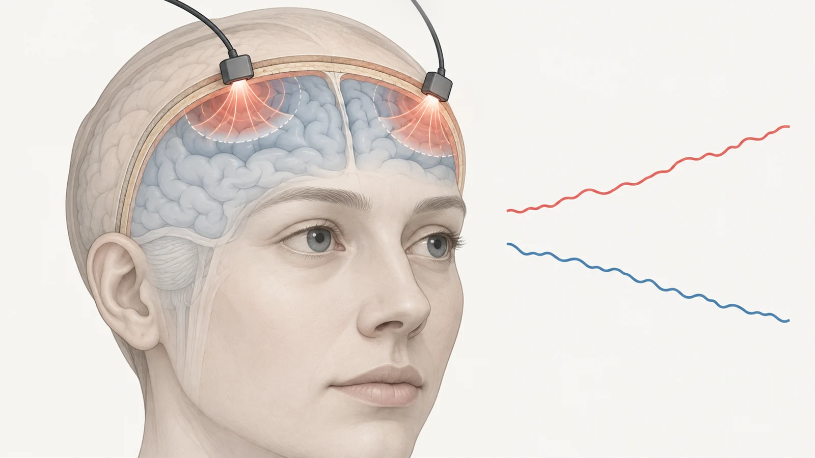
A brief recording from the forehead found a tighter relationship between slow changes in blood volume and a brain-fluid-related signal among people with Alzheimer’s disease than among healthy older adults. The pattern may seem like more coordinated fluid movement. But stronger coupling does not necessarily mean healthier circulation or better waste clearance. It may instead reflect changes in how the brain’s blood and fluid compartments accommodate one another.
The study compared 16 people diagnosed with Alzheimer’s disease, 27 healthy older adults, and 26 healthy young adults. Its findings offer a way to investigate these slow physiological relationships without an invasive procedure. They do not show that the measurement can identify Alzheimer’s in an individual.
Two forehead signals, separated by their optical behavior
The research team used two broadband near-infrared spectroscopy channels placed symmetrically on the forehead. Each sent light into the head and measured light scattered back from the tissue. The recorded wavelengths allowed the researchers to derive changes in total hemoglobin concentration, used as a proxy for changes in cerebral blood volume, and changes in water content associated with relatively free water in the cerebrospinal fluid (CSF), the fluid around the brain.
The measurement was shallow. With the probes used, the light primarily sampled scalp, skull, the CSF space beneath the skull, and superficial tissue in the prefrontal cortex. The inferred CSF-related water fluctuations therefore concern the superficial space near the brain’s surface, not fluid in deep ventricles or deeper brain regions. The method estimates these components from the optical signal; it does not directly observe fluid moving through the brain.
The team analyzed seven-minute recordings. It separated the signals into three slow infraslow frequency bands—endogenic, neurogenic, and myogenic—and calculated both the correlation and the slope between blood-volume and CSF-related water changes. A more negative correlation means that when one signal fluctuated upward, the other tended to fluctuate downward over time. The slope describes how much the water-related signal changed in relation to a change in the blood-volume signal.
Opposing changes are physiologically interesting because blood, brain tissue, and CSF share the limited space inside the skull. A change in one compartment may be accompanied by a change in another. The measured anticorrelation is consistent with that broad volume-compensation idea, but the recording does not reveal the precise route or cause of the shifts.
The Alzheimer’s group had more negative correlations
Compared with healthy older adults, the Alzheimer’s group had significantly more negative blood-volume/CSF-related water correlations in all three frequency bands. The reported effect sizes for these correlation differences ranged from medium to large. For the slopes, group differences were significant in the endogenic and neurogenic bands; the myogenic-band result was marginal, with a reported p value of 0.053.
The older-adult comparison included an age-adjusted analysis. The age effect and the age-by-group interaction were not significant in that model, so the difference between the older groups was not explained by the age variation within those groups in this analysis. The result is a group-level difference in a small sample, not an estimate of how accurately the measure would classify a new patient.
A stronger inverse relationship could arise if a given blood-volume fluctuation is accompanied by a larger CSF-related water fluctuation. That is one reason the authors discuss reduced intracranial compliance—the capacity of the contents of the skull to accommodate changes in volume—as a possible explanation. The study did not directly measure compliance, however, and its optical coupling metric cannot distinguish that account from other changes in slow fluid regulation.
Young adults provide a separate age comparison
The young-adult group had the weakest coupling overall, with coupling tending to strengthen from young adults to healthy older adults and then to the Alzheimer’s group. Within the healthy groups, the age-related increase was significant in the endogenic and myogenic bands, which the authors describe as more directly linked to cerebrovascular dynamics.
For the young-adult comparison, the researchers pooled and averaged correlation and slope values from five separate seven-minute pre-stimulation baseline recordings. They treated those baselines as comparable with the resting recordings from the two older groups. This difference in recording context matters when interpreting the age pattern.
The pattern across groups is compatible with the idea that blood vessels in younger brains buffer volume changes more effectively, while reduced buffering with age could transmit more of a blood-volume swing into the surrounding compartment. The Alzheimer’s group’s stronger coupling might then reflect a further change in that relationship. This remains a proposed physical interpretation, not a mechanism demonstrated by the recordings. The data do not establish whether the stronger coupling is a compensatory response, pathological stiffening, or another form of altered regulation.
What the measurement leaves unresolved
The two-channel setup did not include short-separation probes to isolate signals from scalp and skull. Those superficial tissues can contribute to the optical measurements, so the team could not remove their influence from the reported coupling values. Better separation of superficial and brain-related signals would help determine how much of the observed relationship comes from the compartments the researchers intended to measure.
The Alzheimer’s group included 16 participants. Although the authors described them as being at an early stage, the study did not use objective cognitive assessments to confirm or quantify that staging. The sample size and staging limit how confidently the group result can be generalized, especially to people at different points in the disease or to individuals whose diagnosis is uncertain.
The measured frequency range also matters. The analysis focused on slow oscillations, not the faster cardiac-driven pulsations often considered in discussions of intracranial compliance. And while CSF is involved in brain waste-clearance pathways, this measurement does not directly track glymphatic transport or show how much waste is cleared. It captures a relationship between two inferred, superficial signals; it does not establish diagnostic accuracy, predict disease before symptoms, or demonstrate clinical benefit.
What a forehead reading could add to dementia assessment
A short, noninvasive forehead measurement could eventually give clinicians a physiological measure to consider alongside established assessment of older adults, adding information about slow blood-and-fluid regulation without requiring an invasive CSF procedure. That possibility depends on whether the group pattern is replicated, superficial tissue contributions can be separated more reliably, and the measure proves useful when interpreted with clinical findings. In this study, it remains a promising research measure rather than a test for patients.
Key Sources
- Primary source: Cerebrovascular-CSF coupling measured by broadband near-infrared spectroscopy as a physiological marker of brain aging and Alzheimer's disease.
DOI or Journal Page · Peer-Reviewed Paper · 2026-05-01
Keep exploring