
Research Signal – Source date 2024-05-28
Amyotrophic lateral sclerosis, or ALS, is a neurodegenerative disease in which the upper and lower motor neurons that control movement are progressively lost. The disease can arise in sporadic and genetic forms, and the review published on May 28, 2024, describes survival after onset as commonly limited to two to five years. Its central question is not whether ALS is simply “accelerated aging.” It is whether some biological processes that accumulate with age help create the conditions in which ALS begins or progresses.
One possibility discussed in the review is that cells showing signs of biological aging may alter the environment around motor neurons. If so, drugs designed to remove selected senescent cells could eventually become relevant to movement and independence. But the evidence assembled in this review supports a therapeutic rationale to investigate, not a demonstrated ALS treatment benefit.
Age changes do not follow one simple disease clock
Chronological aging is the passage of time. Biological aging refers to the age-related changes that accumulate in cells and tissues, and those changes do not occur at the same rate in every person or every organ. Genetic background and environmental factors can influence how quickly functional decline appears. This helps explain why people of the same chronological age can have very different biological states.
ALS becomes more common with advancing age, but its age pattern is not identical to those of other neurodegenerative diseases. The review describes ALS risk as very low before age 40, rising after midlife, and appearing to decline in very old adults. Parkinson’s disease emerges around a similar stage and then reaches a plateau in the comparison presented, while Alzheimer’s disease begins later and continues to rise with age.
Those patterns are informative but difficult to interpret as a simple biological timetable. Differences in diagnosis, disease progression, clinical presentation, and the ability to identify cases can distort comparisons between diseases. The broader point is that age may shape ALS risk without making ALS equivalent to ordinary age-related decline. Age-related muscle weakness and loss of motor units overlap with parts of ALS biology, yet the relationship between the two remains more complicated than a single aging switch being turned up.
What patient samples and models reveal about biological aging
The review brings together several reasons to examine biological aging in ALS. Studies of people with ALS have reported a systemic increase in cells carrying senescence-associated features, particularly among patients with bulbar involvement. Muscle biopsies and cultured muscle cells from ALS patients have also shown elevated senescence markers. In motor-cortex tissue, markers such as p16 and p21 have been detected alongside neuronal and astrocyte markers.
These observations place senescence-related biology in tissues affected by ALS, but they do not answer its role in the disease. A marker can indicate that a cell has entered an altered state without showing whether that state initiated the disease, appeared in response to injury, or helped sustain progression. Finding marked cells in patient material also does not show that removing them would improve strength, survival, or another clinical outcome.
The review also discusses damage to DNA and the structures that protect chromosome ends. Spinal tissue from people with C9orf72-associated ALS has shown increased DNA double-strand breaks and markers of the DNA damage response. Postmortem spinal-cord tissue from ALS patients has been reported to have shorter telomeres, repetitive DNA sequences that help protect chromosome ends during cell division.
An experimental example makes the connection more concrete. When SOD1 mice were bred with mice lacking normal telomerase activity, disease onset accelerated and survival fell. That result links age-dependent telomere biology with disease expression in that model. It does not establish that telomere shortening is the initiating cause of ALS in people, but it gives researchers a reason to test how accumulated cellular damage might alter disease vulnerability.
When aging cells change the neighborhood around motor neurons
Cellular senescence is often introduced as a state in which a cell permanently stops dividing. That description captures an important feature, but it becomes harder to apply cleanly in the nervous system. Many neurons are already post-mitotic, meaning they do not normally divide. Researchers therefore look for broader patterns, including regulatory changes and markers such as p16 and p21, as well as changes in the signals a cell releases.
Senescence can serve useful purposes. It can help remove unwanted cells during development and remodeling, and it can limit the uncontrolled growth of damaged or potentially cancerous cells. With persistent DNA damage, shortened telomeres, oxidative stress, or disrupted mitochondria, however, some senescent cells can enter a harmful secretory state.
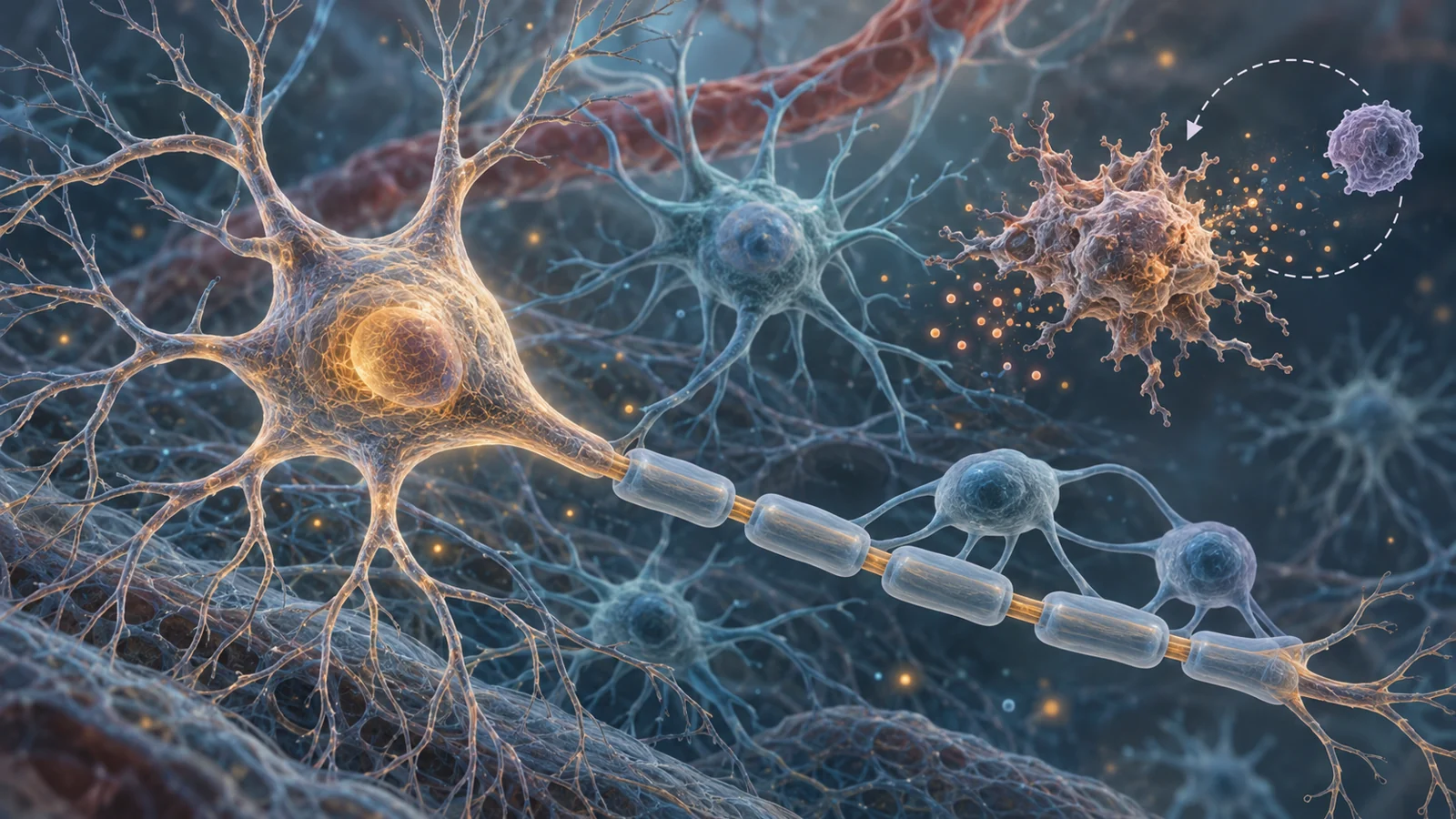
This state is known as the senescence-associated secretory phenotype, or SASP. Instead of remaining biologically quiet, affected cells may release inflammatory cytokines, reactive oxygen species, and other molecules that influence surrounding tissue. The review connects this process with chronic, low-grade inflammation and proposes that it may intensify neuroinflammation in ALS.
Glial cells make the local environment especially important. Microglia and astrocytes normally help protect the central nervous system and regulate the conditions in which neurons function. Evidence from brain and spinal-cord tissues of people with familial ALS associated with SOD1 and C9orf72, and of people with sporadic ALS, suggests that glial senescence may be accelerated in the disease context. Affected glial cells could release inflammatory signals that spread to neighboring glia and other cells, while the loss of normal support may make the environment less favorable for motor neurons.
The proposed mechanism is therefore relational rather than confined to one visibly damaged neuron. A senescence-like state in support cells could influence inflammation, synapses, protein handling, and neuronal survival in the surrounding tissue. The review presents this as a plausible connection between aging biology and ALS pathology, not as proof that every senescent marker has the same meaning or effect.
Why senolytics are being considered
Senolytics are drugs intended to selectively eliminate cells with senescence-like features. Their appeal follows from a simple therapeutic idea: if a persistent population of harmful cells is helping maintain inflammation, reducing that population might improve the tissue environment.
The review describes encouraging findings from aging research. Removing senescent cells in aged mice has improved age-related brain inflammation and aspects of motor and cognitive function. Cell-clearance approaches have also reduced neuroinflammation and improved disease features in models of Alzheimer’s and Parkinson’s disease. Compounds discussed in this area include dasatinib, quercetin, and fisetin, but the broader rationale matters more than any one drug.
These results motivate investigation in ALS because the disease shows overlapping features, including senescence-associated markers, inflammatory signaling, and damage to motor systems. They do not demonstrate that a senolytic will work in ALS. Findings in aged mice or models of other diseases answer different questions from an ALS clinical trial, and the review does not establish an effective ALS regimen, a demonstrated patient benefit, or an acceptable safety profile for this use.
The hardest part is deciding what to remove
Selective clearance is the central biological tradeoff. A useful intervention would need to reach the particular senescent or senescence-like cell populations implicated in ALS while leaving cells whose senescence-related functions remain protective. Removing a cell simply because it carries one marker could be too blunt, especially when neurons, astrocytes, muscle cells, and immune-related cells may occupy different biological states.
Drug specificity is another concern. Many senolytic compounds act on targets shared by other cells and may produce off-target effects. A therapy could therefore reduce an inflammatory signal while also disrupting a useful repair, survival, or tissue-protective function. The relevant question is not only whether senescent cells can be cleared, but whether the right cells can be cleared at the right place and with enough control to avoid creating new harm.
That selectivity problem also changes how success would need to be judged. Lower levels of a senescence marker would not automatically mean better movement. A meaningful treatment would have to connect targeted cell removal with a healthier neural environment and preserved function in people with ALS. Establishing that connection would require ALS-specific clinical evaluation rather than borrowing outcomes from aging or other neurodegenerative conditions.
What targeted cell clearance could mean for daily life with ALS
The patient-facing possibility is straightforward even though the biology is not. If safe, selective removal of harmful senescent cells could preserve muscle strength and motor function in ALS, people might retain more ability to dress, eat, communicate, move through their homes, or manage other daily tasks independently. They might also need less help from caregivers for longer.
That is a conditional prospect, not an observed outcome of the review or of an established ALS therapy. The benefit would depend on identifying the cell populations that matter, avoiding the useful functions of senescence, and showing that the intervention changes function rather than only laboratory markers. The meaningful promise is more control over everyday life for people with ALS, if targeted clearance can eventually meet those biological and safety demands.
Key Sources
- Primary source: Chronological and Biological Aging in Amyotrophic Lateral Sclerosis and the Potential of Senolytic Therapies.
DOI or Journal Page · Review or Meta-Analysis · 2024-05-28
Keep exploring